Fundus Camera: What It Shows About Your Eye Health
An eye test is not only about reading letters from a chart. The inside of your eyes can also provide useful information about your eye health. To see and record this area clearly, eye doctors may use a fundus camera. A fundus camera takes detailed photographs of the back of the eye. These photographs allow eye-care professionals to examine important structures such as the retina, macula, optic disc, and retinal blood vessels. So, what information can these photographs provide to your doctor? Here is a simple explanation. What Is a Fundus Camera?
A fundus camera is a special camera designed to photograph the inside back surface of the eye, which is called the fundus. The fundus includes several important structures involved in vision, including the retina, macula, optic disc and blood vessels. During fundus photography, the patient normally sits in front of the camera and looks at a target while an image is taken through the pupil. The camera may produce a bright flash for a moment. Depending on the type of camera and the purpose of the examination, the doctor may sometimes use eye drops to dilate the pupil before taking photographs. What Parts of the Eye Can a Fundus Camera Show?
A fundus photograph provides doctors with a clear visual record of several structures at the back of the eye.
The Retina
The retina is a thin layer of light-sensitive tissue located at the back of the eye. It receives light and transmits visual information to the brain. Through retinal imaging, doctors can examine the appearance of the retina and search for unusual changes such as bleeding, deposits, swelling, or abnormal blood vessels. Not every retinal change causes noticeable symptoms immediately. This is one of the reasons why retinal examinations can be beneficial in eye care.
The Macula
The macula is a small space at the center of the retina. It is responsible for much of the detailed central vision that we use for activities such as reading, recognizing faces and seeing fine details. A fundus photograph allows the doctor to examine the visible appearance of the macula. Changes in this area may be associated with conditions such as age-related macular degeneration or diabetic eye disease. If a closer look at the retinal layers is needed, the doctor may recommend additional imaging such as OCT.
The Optic Disc
The nerve that leads to the retina is joined to the optic disc. From your eye to your brain, the optic nerve sends messages about what you see. With optic nerve imaging, doctors can document the appearance of the optic disc and compare it with photographs taken during later examinations. Changes in the appearance of the optic nerve may sometimes require further investigation for conditions such as glaucoma. A fundus photograph alone, however, does not diagnose glaucoma. Other examinations may also be required.
Why Are Retinal Blood Vessels Important in an Eye Exam?
One interesting feature of the retina is that its blood vessels can be directly viewed and photographed. A fundus eye examination allows doctors to observe the appearance and pattern of these vessels. Certain changes in retinal blood vessels can occur in people with conditions such as diabetes or high blood pressure. For example, doctors may notice bleeding, changes in vessel appearance, or other abnormalities. These findings do not automatically mean that a person has a particular disease. They need to be interpreted along with medical history and other examinations. Can Fundus Photography Help Find Diabetic Eye Changes?
Yes. Diabetic retinopathy screening is an important use of retinal photography. Diabetes can affect the small blood vessels inside the retina. During the early stages, a person may not notice any obvious change in vision. Fundus photographs can help trained eye-care professionals identify and document visible retinal changes associated with diabetic retinopathy. Regular eye examinations are therefore particularly important for people with diabetes, even when their eyesight appears normal.
Why Do Doctors Keep Fundus Photographs?
A useful feature of fundus photography is that the image can become part of a patient's eye-health record. Suppose a photograph is taken today and another is taken during a future examination. The doctor can compare the two images. This comparison may help identify whether an area has remained stable or changed over time. For example, doctors may compare changes in the retina, optic disc, macula or blood vessels. This makes fundus photographs useful not only for examining the eye at one point in time but also for monitoring certain changes during future visits.
Fundus Camera vs OCT: Are They the Same?
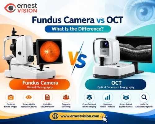
A fundus camera and OCT are both used to examine the back of the eye, but they provide different types of information. A fundus camera produces a photograph showing the visible appearance of the retina, optic disc, macula and blood vessels. OCT, or optical coherence tomography, provides detailed cross-sectional images of the retinal layers. It allows doctors to examine structures beneath the visible retinal surface in greater detail. Because they provide different information, doctors may sometimes use both tests as part of an eye examination. Is Fundus Photography Painful?
Fundus photography is generally a non-invasive procedure. The patient usually places their chin and forehead against the supports of the camera and looks at a target. The camera then takes one or more photographs. You may notice a bright flash, but taking the photograph itself is usually quick. If pupil-dilating drops are required, your vision may temporarily become blurred or more sensitive to light afterward. Your eye-care professional can explain what to expect.
Can a Fundus Camera Detect Every Eye Problem?
No. A fundus photograph provides useful information about the back of the eye, but it cannot detect every eye condition. Some problems involve other structures, such as the cornea or lens, while others require more detailed testing. Depending on the findings and symptoms, an eye-care professional may also use a slit lamp, tonometer, visual field test, OCT or other diagnostic equipment. Therefore, retinal photography should be considered one part of a complete eye examination rather than a replacement for one.
Why Is Retinal Imaging Important Even When Vision Seems Normal?
Good vision does not always mean that every part of the eye is healthy. Some retinal conditions may develop gradually and cause few noticeable symptoms in their early stages.
Retinal imaging gives eye-care professionals a way to examine and document structures that you cannot see yourself. It can also create a useful baseline image. Future photographs can then be compared with earlier ones if your doctor needs to monitor changes. For patients, understanding this purpose can make a retina examination easier to understand and less unfamiliar.
Frequently Asked Questions
What is a fundus camera used for?
A fundus camera is used to photograph structures at the back of the eye, including the retina, macula, optic disc and retinal blood vessels.
What can fundus photography detect?
Fundus photography can show visible retinal and optic-disc changes that may be associated with diabetic retinopathy, macular degeneration, retinal vascular problems and some optic nerve conditions. Additional tests may be required for diagnosis.
Can a fundus camera detect diabetic retinopathy?
A fundus camera can photograph retinal changes associated with diabetic retinopathy. Diabetic retinopathy screening commonly uses retinal photographs to help identify people who may need further eye assessment.
Can a fundus camera detect glaucoma?
A fundus photograph can show and document the appearance of the optic disc. Certain changes may raise concern about glaucoma, but a fundus image alone cannot confirm a glaucoma diagnosis.
Does fundus photography hurt?
No. Fundus photography is generally non-invasive and quick. A bright flash may be noticeable when the photograph is taken.
Do your eyes need to be dilated for fundus photography?
Not always. Some fundus cameras can take useful images without pupil dilation. In other cases, dilating drops may be used to provide a clearer or wider view.
What is the difference between a fundus camera and OCT?
A fundus camera photographs the visible surface of the retina and related structures. OCT creates detailed cross-sectional images of retinal layers. The two tests provide different but complementary information.