

Understand fundus camera vs OCT, their imaging methods, features, benefits and differences. A practical guide for eye clinics and hospitals in Dubai.
Fundus Camera vs OCT: Differences, Uses & Benefits
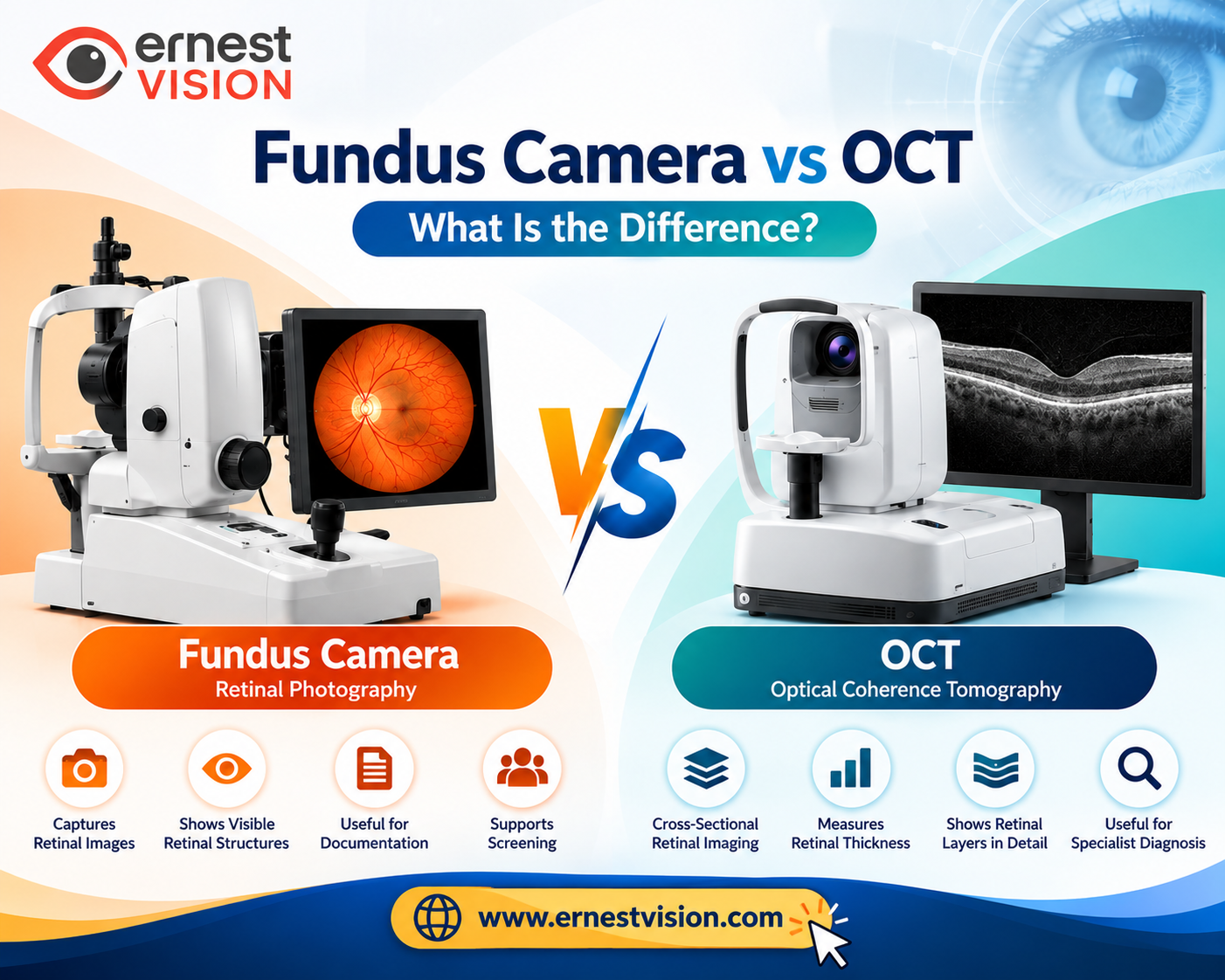
When an eye specialist needs to examine the retina, two commonly used imaging instruments are a fundus camera and an Optical Coherence Tomography (OCT) machine. Both help doctors study the back of the eye, but they do not produce the same type of image. The main difference in Fundus Camera vs OCT is how the retina is examined. A fundus camera takes a photograph showing the visible retinal surface, while an OCT machine produces detailed cross-sectional images of the retinal layers.
For ophthalmologists, optometrists, eye hospitals and clinic owners in Dubai, understanding these differences is important when planning retinal imaging facilities. The right equipment depends on the examinations performed, the type of patients seen and the level of detail required.
What Is a Fundus Camera?
A Fundus Camera is a specialised medical imaging instrument used to photograph the inside surface of the eye. It captures images of important structures, including the retina, macula, optic disc and retinal blood vessels. During an examination, the patient looks towards a fixation target while the camera captures an image of the retina. The photograph can then be viewed on a computer, saved in the patient's medical record or compared with images from previous visits. Fundus photography is commonly used for retinal documentation and screening. It allows eye specialists to observe visible changes in the retina without needing to draw or manually record every detail.
Important Features of a Fundus Camera
Different models offer different capabilities, but commonly available features include:
- High-resolution digital retinal photography
- Colour images of the retina
- Automatic or manual focusing
- Retinal image storage and viewing software
- Non-mydriatic imaging in supported models
- Different fields of view depending on the camera
- Image export for patient records
Some instruments also provide wide-field photography, autofluorescence or other imaging modes.
Benefits of Using a Fundus Camera
A fundus camera is useful when an eye clinic needs clear photographic documentation of the retina. It helps clinicians compare retinal appearance over time, identify visible abnormalities and explain findings to patients using actual images. Fundus photography also plays an important role in diabetic eye screening programmes. However, a standard fundus photograph does not directly measure retinal thickness or display individual retinal layers.
What Is an OCT Machine?
Optical Coherence Tomography (OCT) is an imaging technology that uses light to produce detailed cross-sectional pictures of structures inside the eye. Unlike a fundus camera, which mainly records the visible surface, OCT provides information about the internal layers of the retina. An OCT examination can help doctors assess retinal thickness, macular structure and changes affecting the retinal nerve fibre layer. The scan is non-invasive and usually does not require physical contact with the eye.
Important Features of an OCT Machine
Depending on the manufacturer and model, an OCT system may include:
- High-resolution retinal cross-sectional imaging
- Retinal thickness measurement
- Macular thickness maps
- Retinal nerve fibre layer analysis
- Optic nerve head assessment
- Three-dimensional retinal imaging
- Scan comparison and progression analysis
- OCT angiography or anterior segment imaging in supported systems
These functions are especially useful in specialist retina and glaucoma services.
Benefits of OCT Imaging
OCT helps eye specialists examine structural changes that may not be clearly visible in ordinary retinal photographs. For example, it can help identify fluid within the retina, changes in macular thickness or abnormalities affecting retinal layers. It also provides measurements that can be compared during follow-up examinations. OCT findings must still be interpreted alongside the patient's symptoms, clinical examination and other diagnostic tests.
Fundus Camera vs OCT: Key Differences
The two instruments have different imaging methods, outputs and clinical applications.
| Comparison | Fundus Camera | OCT Machine |
|---|---|---|
| Imaging technology | Digital retinal photography | Optical coherence tomography |
| Image type | Two-dimensional retinal photograph | Cross-sectional and three-dimensional retinal images |
| Main purpose | Viewing and documenting visible retinal structures | Examining internal retinal layers |
| Retinal thickness measurement | Not available with standard fundus photography | Available in suitable scan protocols |
| Optic disc imaging | Records visible optic disc appearance | Provides structural measurements of the optic nerve region |
| Diabetic eye assessment | Documents visible retinal changes | Helps assess diabetic macular oedema |
| Glaucoma assessment | Supports optic disc documentation | Measures structures such as retinal nerve fibre layer thickness |
| Image storage | Digital photographs | Scans, measurements and reports |
| Common clinical setting | General ophthalmology and screening | Retina, glaucoma and specialist diagnostic services |
The difference is not simply image quality. Each instrument provides a different kind of clinical information.
How Does a Fundus Camera Work?
A Digital Fundus Camera uses illumination and an optical imaging system to capture a photograph of the retina. The patient is positioned in front of the instrument, usually with the chin and forehead supported. The operator aligns the camera and asks the patient to look towards a fixation target. Once the image is captured, it appears on the display for review. The photograph shows the appearance of retinal blood vessels, the macula and the optic nerve head. The quality of the image can depend on several factors, including pupil size, eye movement and the clarity of the eye's optical media. Some fundus cameras can capture images without routine pupil dilation, while others may require dilation depending on the examination.
How Does an OCT Machine Work?
An OCT Machine uses low-power light and interferometry to measure reflections from different depths within eye tissue. The system processes these measurements to produce cross-sectional images of the retina. During the examination, the patient looks at a fixation target while the device scans the required area. The resulting images allow the clinician to examine retinal layers and assess changes in thickness or structure. OCT is particularly helpful when a condition affects tissue beneath the visible retinal surface.
Clinical Applications of Fundus Camera and OCT
Both instruments are used in ophthalmology, although their roles vary according to the condition being investigated.
1. Diabetic Retinopathy
Fundus photography helps document visible retinal changes such as haemorrhages, microaneurysms and exudates. OCT is particularly useful when assessing diabetic macular oedema because it can show fluid accumulation and changes in retinal thickness.
2. Glaucoma Assessment
A fundus camera provides photographs of the optic disc, helping clinicians document its appearance. OCT provides measurements of structures such as the retinal nerve fibre layer and ganglion cell complex, depending on the system. These findings are used alongside intraocular pressure measurements, visual field testing and clinical examination.
3. Age-Related Macular Degeneration
Fundus photographs can show visible macular changes, including drusen and pigment abnormalities. OCT can provide additional information about retinal structure and fluid associated with certain forms of macular degeneration.
4. Retinal Documentation
Fundus photography is particularly useful for recording the appearance of retinal structures over time. OCT can supplement this information when a more detailed structural examination is needed.
Can a Fundus Camera Replace OCT?
A standard fundus camera cannot completely replace an OCT machine because the two devices provide different information. A fundus photograph can show visible changes on the retinal surface, but it cannot provide the same detailed cross-sectional view of retinal tissue. Similarly, although some OCT systems include fundus-like reference images or additional imaging features, these are not always equivalent to dedicated colour fundus photography. In many ophthalmology practices, the two technologies are used together. For example, a clinician may review a fundus photograph to observe the overall retinal appearance and then use OCT to examine a specific area of the macula in greater detail.
Which Equipment Is Suitable for an Eye Clinic in Dubai?
Choosing between a fundus camera and OCT depends on the services offered by the clinic. A general eye examination centre may need retinal photography for routine documentation and screening. A specialist retina or glaucoma clinic may require OCT for detailed structural imaging. Larger ophthalmology centres may benefit from both technologies. Before selecting Retinal Imaging Equipment, clinic managers should consider the following factors.
Clinical Services
Determine whether the clinic focuses on general eye examinations, diabetic retinopathy screening, glaucoma care or specialist retinal services.
Imaging Requirements
Check whether the clinical team needs colour retinal photography, cross-sectional scans, thickness measurements or additional imaging modes.
Patient Volume
Consider the expected number of examinations and how easily the equipment fits into the clinic's daily workflow.
Software Compatibility
Review image storage, reporting functions and compatibility with existing electronic medical record systems.
Training and Maintenance
Check the availability of operator training, warranty coverage, servicing and technical assistance.
Regulatory Requirements
Eye clinics and healthcare procurement teams in Dubai should verify applicable UAE medical-device regulatory requirements, supplier documentation and relevant product approvals before procurement.
Important Specifications to Compare Before Purchasing
Product specifications should be reviewed carefully because not every fundus camera or OCT system offers the same capabilities.
| Specification | What to Check |
|---|---|
| Image resolution | Clarity and detail of the captured images |
| Field of view | Area of the retina visible in one photograph |
| OCT scan speed | Number of A-scans captured per second |
| OCT scan resolution | Level of structural detail available |
| Pupil requirements | Minimum pupil size needed for imaging |
| Imaging modes | Colour, autofluorescence, OCTA or other supported functions |
| Software | Image review, measurements and reporting |
| Connectivity | Image export and clinic system integration |
| Installation | Space, power and workstation requirements |
| Support | Warranty, servicing and training |
For OCT equipment, scan speed and axial resolution are particularly important technical specifications. For fundus cameras, field of view, image resolution and focusing capabilities are common points of comparison. Actual specifications should always be confirmed using the manufacturer's technical documentation.
Fundus Camera and OCT Price Considerations in Dubai
The cost of ophthalmic imaging equipment varies according to the brand, model, specifications and included software. A basic digital fundus camera and a wide-field retinal imaging system may have significantly different prices. Similarly, an OCT machine with additional features such as OCT angiography may cost more than a system designed primarily for standard retinal scans. When comparing quotations, buyers should check whether installation, training, warranty, and software licences are included. Maintenance expenses and technical support arrangements also affect the overall cost of ownership. For hospitals and eye clinics, choosing equipment based on clinical requirements and long-term usability is generally more practical than comparing purchase prices alone.
Fundus Camera vs OCT: Which Should You Choose?
The choice depends on the type of retinal information the clinic needs. A fundus camera is suitable when the main requirement is photographing and documenting visible retinal structures. OCT is useful when the examination requires detailed information about retinal layers, macular thickness or optic nerve-related structural measurements. For comprehensive retinal assessment, access to both instruments may be beneficial. Rather than treating them as competing technologies, it is more useful to understand how each contributes to patient examination and clinical documentation.
Frequently Asked Questions
1. What is the main difference between a fundus camera and OCT?
A fundus camera captures photographs of the retina, while OCT produces cross-sectional images showing the internal retinal layers.
2. Is OCT better than a fundus camera?
Neither instrument is better for every examination. OCT provides detailed retinal structural information, while fundus photography is useful for documenting visible retinal features.
3. Can a fundus camera detect diabetic retinopathy?
3. Can a fundus camera detect diabetic retinopathy?
Fundus photographs can show visible retinal changes associated with diabetic retinopathy. A trained professional must interpret the images, and additional examinations may be necessary.
4. Is OCT used for glaucoma diagnosis?
4. Is OCT used for glaucoma diagnosis?
OCT helps assess structural changes associated with glaucoma, including retinal nerve fibre layer measurements. It is one part of a broader glaucoma evaluation.
5. Does a fundus camera require pupil dilation?
Not always. Some non-mydriatic cameras can capture retinal photographs without routine dilation, although dilation may be needed in certain patients.
6. Are fundus photography and OCT safe?
Both are generally considered safe, non-invasive imaging examinations. Neither uses ionising radiation.
7. Can an eye clinic use both fundus photography and OCT?
Yes. Many clinics use both because the images provide complementary information about retinal health.
8. What factors affect OCT machine prices in Dubai?
Pricing depends on the manufacturer, scan technology, imaging features, software, warranty, installation and technical support arrangements.
Conclusion
Understanding the difference between a fundus camera and OCT helps eye care professionals select equipment according to their clinical needs. A fundus camera provides photographs of visible retinal structures, while OCT offers detailed images of the layers within the retina. Both technologies play important roles in retinal examination, disease assessment and patient follow-up. For ophthalmologists, eye hospitals and healthcare procurement teams in Dubai, comparing clinical applications, imaging features, software and technical support is an important part of equipment selection. Ernest Vision provides information about ophthalmic instruments and diagnostic equipment for eye care professionals and healthcare organisations.