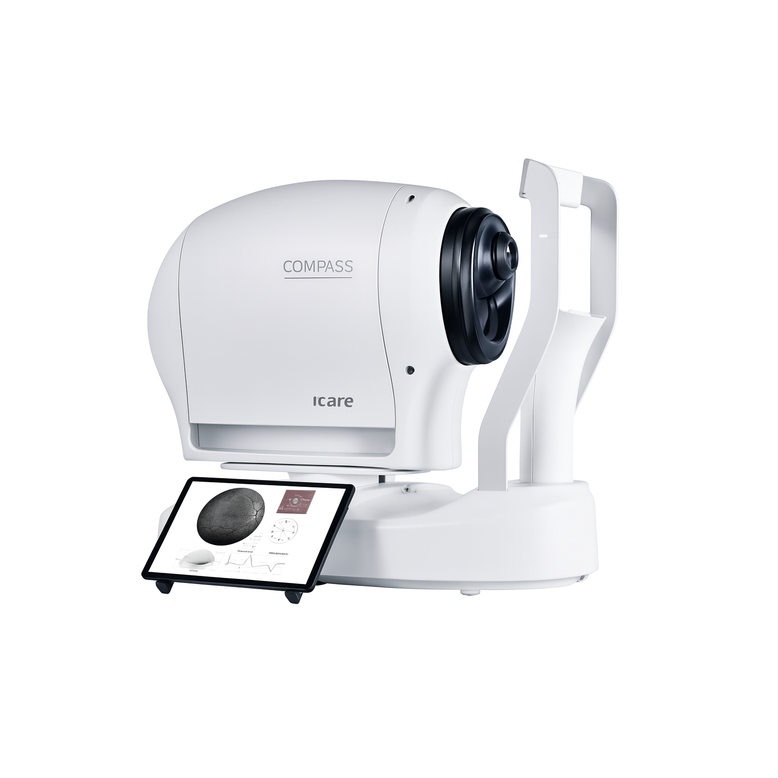
The iCare COMPASS is a breakthrough innovation in ophthalmic diagnostics, combining fundus-guided perimetry, TrueColor confocal retinal imaging, and automated visual field testing into a single, compact device. Built for modern glaucoma and retinal clinics, COMPASS sets a new global benchmark in functional eye assessment, delivering unmatched precision, repeatability, and real-time structure–function correlation.
At its core, COMPASS features a high-resolution TrueColor confocal retinal camera that captures sharp, low-noise fundus images simultaneously with the visual field exam. These structural images allow clinicians to visually correlate glaucomatous damage, macular disorders, and neuro-ophthalmic abnormalities with functional deficits—providing a comprehensive diagnostic picture that enhances decision making and patient management.
The system supports a wide range of perimetry strategies including Full Threshold, Screening, Fast Thresholding, and Progression Analysis. Its intuitive user interface, ergonomic design, and automated workflow make it ideal for high-volume clinical environments. With enhanced patient comfort, minimal training requirements, and superior test reproducibility, COMPASS elevates clinical efficiency and diagnostic confidence.
Powered by Ernest Pharmaceutical Group
(All brands under Ernest Pharmaceutical Pvt. Ltd.)
@ERNESTVISION | ERNEST PHARMACEUTICAL PVT LTD
For global exports, OEM branding & distributor partnerships:
📧 Email: exports@ernestpharmaceuticals.com 🌐 https://ernestpharmaceuticals.com/ | https://www.ernestvision.com/ | https://www.ernestimpex.com/
Sachin Kashiwar: WhatsApp link https://wa.me/919359902383
📲 WhatsApp: +91 9359902383
🔍 Key Clinical Advantages
Eye-Tracking Guided Perimetry
Eliminates fixation losses, reduces artifacts, and improves test repeatability.TrueColor Confocal Fundus Imaging
Provides clear, high-quality retinal images correlated with functional test points.Structure–Function Integration
Enables clinicians to compare visual field defects with corresponding retinal anatomy.Advanced Glaucoma Progression Tools
Ideal for early detection, monitoring, and long-term management of glaucoma patients.Efficient Testing Workflow
Automated alignment, fast acquisition, and user-friendly software reduce exam time.Patient-Friendly Experience
Compact design, quick tests, and improved fixation tracking enhance patient compliance.
⚙️ Technical Advantages
Confocal imaging for high-contrast fundus photography
Automated real-time fixation tracking (live compensation)
Multiple perimetry patterns (24-2, 30-2, 10-2 and custom)
High-resolution retinal imaging for precise pathology mapping
Reliable for glaucoma, macular degeneration, diabetic eye disease & neuro-ophthalmology
Compact footprint suitable for all clinic sizes
EMR-compatible reporting and digital workflow support
🎯 Ideal For
Glaucoma specialists
Comprehensive ophthalmologists
Retina specialists
Eye hospitals & teaching institutes
High-volume diagnostic centers