

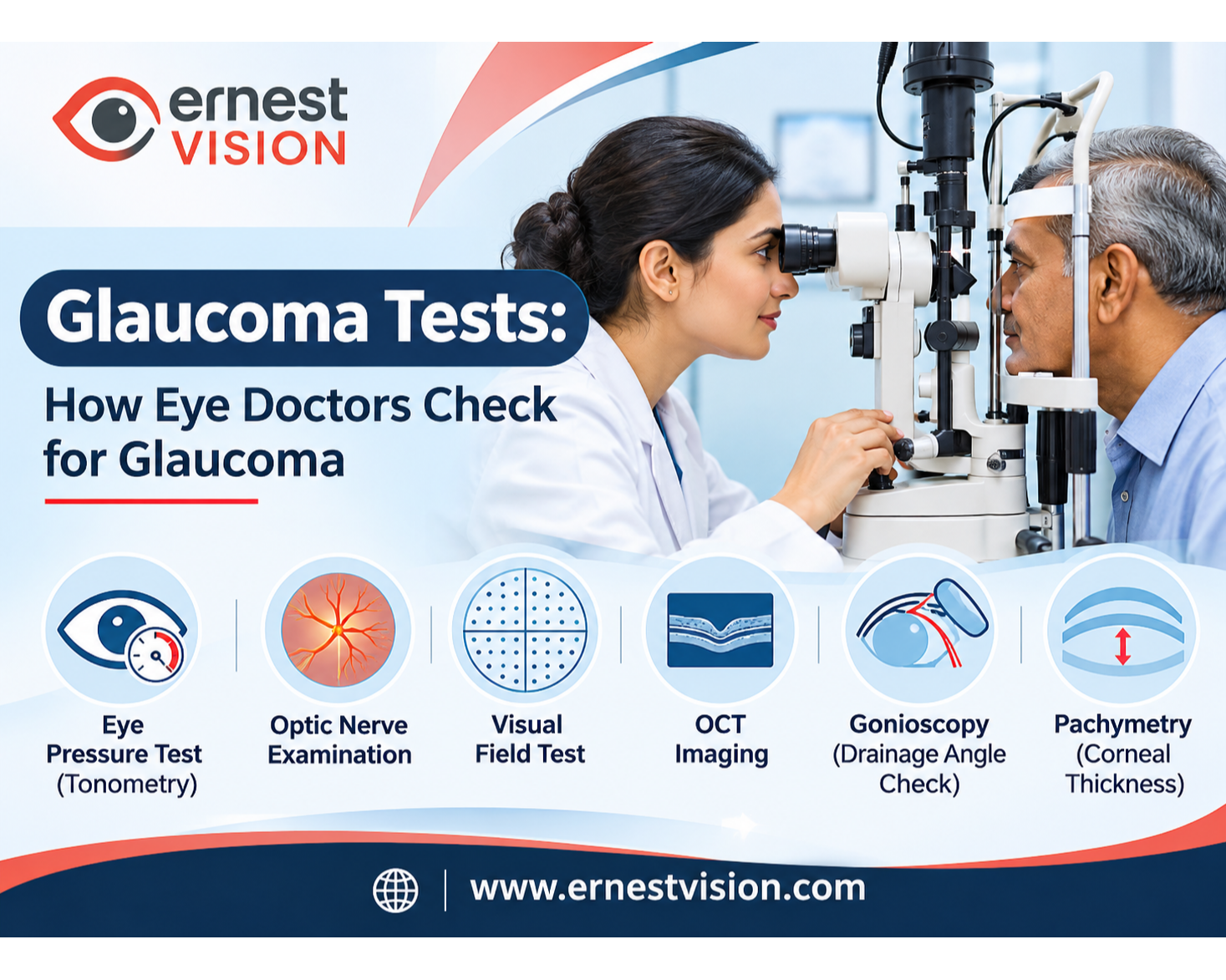
Glaucoma Tests: How Eye Doctors Check for Glaucoma
A glaucoma test involves several eye examinations that help doctors assess the optic nerve, measure pressure inside the eye, and identify possible changes in vision. Glaucoma is a group of eye conditions that can damage the optic nerve, sometimes without noticeable warning signs. There is no single examination that can identify every case of glaucoma. Eye doctors combine different test results with a patient's medical history and individual risk factors to understand eye health and determine whether further evaluation is necessary.
Why Are Glaucoma Tests Important?
Glaucoma can develop slowly, particularly in its common open-angle form. Many people experience no obvious glaucoma symptoms during the early stages. As the disease progresses, changes may affect peripheral vision. Because lost vision generally cannot be restored, identifying the condition and arranging appropriate medical care early is important. According to the National Eye Institute's glaucoma information, a comprehensive dilated eye examination is important for detecting glaucoma. People with a family history of glaucoma, increasing age or other relevant risk factors may need more frequent examinations. An eye-care professional can recommend a suitable schedule.
1. Eye Pressure Test: Measuring Intraocular Pressure
An eye pressure test is a common part of glaucoma evaluation. It measures the pressure inside the eye, medically known as intraocular pressure (IOP). Eye pressure is measured using a device called a tonometer. Different types of tonometers use different measurement methods.
Non-Contact Tonometry
Non-contact tonometry uses a brief puff of air directed toward the cornea. The instrument estimates eye pressure by analyzing the cornea's response.
The test does not require the measuring instrument to touch the eye. It is often used during routine eye examinations and preliminary pressure assessments. Clinics evaluating this equipment can also read about Non-Contact Tonometers for Eye Clinics.
Applanation Tonometry
Applanation tonometry measures the force required to flatten a small area of the cornea. Goldmann applanation tonometry is an established reference method for measuring IOP in clinical practice. It commonly involves anesthetic eye drops and a measuring instrument used with a slit lamp.
For a device-focused explanation, see Ernest Vision's Tonometer AT900 overview.
Rebound Tonometry
Rebound tonometry uses a small probe that briefly touches the corneal surface. The device estimates pressure from the probe's movement.
Some rebound tonometers are portable, making them useful in different clinical examination settings.
The appropriate instrument depends on clinical requirements, the patient's condition, and the equipment available.
2. Optic Nerve Examination
The optic nerve carries visual information from the retina to the brain. Glaucoma can damage this nerve, making optic nerve examination an important part of diagnosis. An ophthalmologist may use a slit lamp with an appropriate examination lens, an ophthalmoscope, or retinal imaging equipment.
What Does the Doctor Look For?
The doctor evaluates the appearance of the optic disc and surrounding nerve tissue. Changes in the optic nerve's structure may suggest glaucoma-related damage. The examination may include dilating eye drops to provide a clearer view of the structures inside the eye. However, optic nerve appearance varies between individuals. A suspicious finding usually needs to be interpreted alongside other examination results.
3. Visual Field Testing: Checking Peripheral Vision
A visual field test evaluates how well a person can detect visual signals in different areas of their field of vision. This is particularly relevant because glaucoma may affect side vision before a person notices difficulties with everyday activities.
How Is a Visual Field Test Performed?
During automated visual field testing, the patient looks at a fixed point inside an instrument called a perimeter. Small lights appear at different locations. The patient responds when a light is visible, and the instrument records the results. The doctor then examines the report for areas of reduced visual sensitivity. Test results can be influenced by concentration, tiredness and familiarity with the procedure. Repeating a visual field test may help determine whether a finding is consistent.
4. OCT Imaging: Evaluating the Optic Nerve
Optical coherence tomography (OCT) is a non-invasive imaging technique that produces detailed cross-sectional images of eye structures. During glaucoma evaluation, OCT can help doctors assess the retinal nerve fiber layer and other structures associated with the optic nerve.
Why Is OCT Useful?
OCT provides measurements and images that may help identify structural changes. When previous images are available, doctors can compare examinations over time to assess whether changes have occurred. An OCT result alone does not confirm or exclude glaucoma. The findings must be considered alongside the optic nerve examination, pressure measurements, and visual field testing.
5. Gonioscopy: Examining the Drainage Angle
Gonioscopy helps an ophthalmologist examine the angle between the iris and cornea. This region contains structures involved in the drainage of fluid from the eye.
How Is Gonioscopy Performed?
The doctor generally applies numbing eye drops before placing a specialized examination lens against the eye. Using the lens and appropriate illumination, the doctor examines the drainage angle. Gonioscopy helps distinguish open-angle from angle-closure conditions and can provide information about other abnormalities affecting the angle. The examination is particularly important when the doctor suspects an angle-related cause of elevated eye pressure.
6. Pachymetry: Measuring Corneal Thickness
Pachymetry measures the thickness of the cornea, the transparent front surface of the eye. Corneal thickness matters because it can affect the interpretation of certain intraocular pressure measurements. For example, a pressure reading may be influenced by the cornea's physical characteristics rather than reflecting pressure alone. Pachymetry therefore provides useful additional information when an ophthalmologist evaluates glaucoma risk. It does not diagnose glaucoma independently.
Which Instruments Are Used in Glaucoma Testing?
Different diagnostic instruments provide different information about the eye.
| Examination | Instrument | Purpose |
|---|---|---|
| Eye pressure measurement | Tonometer | Measures pressure inside the eye |
| Optic nerve assessment | Slit lamp with examination lens or ophthalmoscope | Examines optic nerve appearance |
| Peripheral vision assessment | Visual field analyzer | Measures visual sensitivity |
| Retinal and optic nerve imaging | OCT system | Examines structural features |
| Drainage angle examination | Gonioscopy lens | Evaluates the anterior chamber angle |
| Corneal thickness measurement | Pachymeter | Measures corneal thickness |
These instruments serve different clinical purposes. Not every person requires every test during each appointment.
Eye clinics planning examination facilities may also find the Complete Ophthalmic Equipment Overview useful for understanding the broader categories of diagnostic instruments.
The American Academy of Ophthalmology's Glaucoma Summary Benchmarks describe the clinical examinations and diagnostic methods considered during glaucoma evaluation.
What Is Normal Eye Pressure?
Normal eye pressure is a common topic during glaucoma examinations. Eye pressure is expressed in millimeters of mercury (mmHg). A range of approximately 10–21 mmHg is commonly used as a reference, but these numbers are not a definitive diagnostic cutoff. Some people have pressure above the usual reference range without detectable glaucoma damage. Others develop glaucoma despite measurements within that range. The National Eye Institute explains in its guidance on glaucoma detection that tonometry alone is insufficient for an accurate diagnosis. This is why ophthalmologists consider optic nerve health, visual function, corneal thickness, and other clinical findings rather than relying on a single reading.
What Happens After a Glaucoma Examination?
Once the tests are completed, the ophthalmologist reviews the findings together. If there are no suspicious changes, the doctor may recommend routine follow-up based on the person's risk factors. If the examination identifies uncertain findings, additional testing or monitoring may be recommended. Repeated measurements can help doctors assess changes over time. A person with confirmed glaucoma may require ongoing care to reduce the risk of further optic nerve damage. The testing and follow-up plan depends on the individual's clinical situation.
Conclusion
A glaucoma test is best understood as a combination of examinations rather than one procedure. An eye pressure test provides important information, but optic nerve assessment, visual field testing, OCT imaging, gonioscopy and pachymetry may also contribute to a complete evaluation.
These tests help ophthalmologists identify suspicious findings, investigate glaucoma and monitor eye health over time. Because early glaucoma symptoms may be absent, professional eye examinations remain important for people with known risk factors and those advised to undergo regular screening.
Frequently Asked Questions
1. What is a glaucoma test?
A glaucoma test refers to one or more examinations used to assess optic nerve health, eye pressure and visual function. Doctors often combine several methods when investigating glaucoma.
2. Is a glaucoma test painful?
Most routine glaucoma examinations cause little or no discomfort. Some contact-based procedures use numbing eye drops. The experience varies depending on the test.
3. Can an eye pressure test detect glaucoma?
An eye pressure test helps identify elevated pressure, an important glaucoma risk factor. However, pressure measurement alone cannot confirm or rule out glaucoma.
4. What is normal eye pressure?
Normal eye pressure is commonly described using a reference range of approximately 10–21 mmHg. Individual interpretation depends on optic nerve findings, corneal characteristics and other clinical factors.
5. Why do doctors perform visual field tests?
Visual field tests help identify changes in peripheral vision that may be associated with glaucoma.
6. What is the purpose of OCT in glaucoma testing?
OCT provides detailed images of retinal and optic nerve structures. Doctors may use these images to assess changes and monitor findings over time.
7. Can glaucoma occur without symptoms?
Yes. Many people experience no noticeable glaucoma symptoms during the early stages, especially with open-angle glaucoma.
8. How often should someone have a glaucoma examination?
The recommended frequency depends on age, family history, eye health and other risk factors. An eye-care professional can recommend an appropriate examination schedule.Medical Disclaimer: This article is intended for general educational purposes. It does not provide an individual diagnosis or treatment recommendation. Sudden severe eye pain, redness, blurred vision or nausea associated with eye symptoms requires urgent medical attention.