Thank you for writing to us. One of our executive will reach back to you through your submitted medium. In case there’s an urgency, feel free to connect over WhatsApp for faster response.
Prefer calling? Dial +919359902383 (International callers) or +919359902383 (Indian callers).
Product introduction
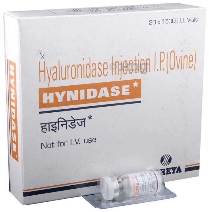
Hynidase Injection is used as a preservative in combination with other medicines. It enhances the absorption and spread of fluids for hydration, improves the dispersion and effectiveness of injected drugs, and improves the absorption of contrast agents used in certain imaging tests, like subcutaneous urography.
Generally, Hynidase Injection comes in combination with injectable medicines. It must be administered as per prescription under the supervision of a doctor or a nurse. Let your doctor know if you have any known allergies to this medication.
Hynidase Injection is a safe medicine and has minimal or no side effects. However, some patients might experience injection site reactions such as redness, swelling, and pain. Let your doctor know if they bother you.
Marketer
Shreya Life Sciences Pvt Ltd
SALT COMPOSITION
Hyaluronidase (1500IU)
Storage
Store below 30°C
Uses of Hynidase Injection
Preservative
Benefits of Hynidase Injection
In Preservative
Hynidase Injection is used as a preservative to prevent damage to the original form of the medicine that is given as an injection by a doctor or nurse. If it is not present, harmful organisms can contaminate the solution and cause infections or reactions in the person who takes any such medicine. It also acts as a solvent for certain medicines and helps in better absorption of the medicine in our body.
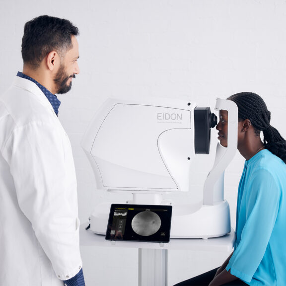
iCare EIDON — the first TrueColor Confocal imaging device for ultra-high resolution widefield imaging
iCare EIDON Family combines a confocal optical engine with a white light LED source – which includes the entire visible spectrum of light – to illuminate the retina and to capture TrueColor fundus images characterized by colors close to reality. It’s the perfect retinal imaging system that provides TrueColor and widefield views in multiple imaging modalities. iCare EIDON provides unsurpassed image quality and a unique, live, confocal view of the retina in a dilation-free operation.
Key features
Multiple imaging modalities including TrueColor, blue, red and Red-Free and infrared confocal images
Widefield, ultra-high-resolution imaging
Capability to image through cataract and media opacities
Dilation-free operation (minimum pupil 2.5 mm)
Flexibility of fully automated and fully manual mode
All-in-one compact design, no additional PC required
Fundus Imaging System Features
Non-mydriatic
minimum pupil size 2.5 mm
Field of view (measured from the center of the eye)
Individual image
90°
Mosaic (up to nine images)
up to 160°
Sensor resolution
14 Mpixel (4608 x 3288)
Light sources
White LED
440 - 650 nm
Infrared LED
825 - 870 nm
Imaging modalities
TrueColor
Infrared
RGB channels separation*
Other features
Working distance
28 mm
Offering the best of confocal technology, iCare EIDON guarantees increased image sharpness, better optical resolution, higher details and greater contrast that takes retinal diagnostics to the next level. This combined with its easy-to-use interface and patient-friendly features make the iCare EIDON a valuable and efficient tool in any clinical setting.
“EIDON TrueColor Widefield Confocal Scanner gives great retinal images, far superior to a standard retinal camera: you are able to see certain condition in much better visualization, such as Diabetic Retinopathy.” – Steven G Ferrucci, OD, FAAO, US
Ultra-Widefield details in high definition
iCare EIDON’s widefield optics from 90° to 160° field of view allows imaging of the central retina as well as the periphery. iCare EIDON TrueColor confocal technology applied to widefield imaging is particularly crucial as it helps to improve the detection, analysis and monitoring of pathologies that could arise in the periphery of the retina. iCare EIDON’s widefield features help to preserve the sharpness and details even in the periphery, facilitating improved and early diagnosis.
Increased field of view up to 200˚
Thanks to the EIDON Ultra-Widefield Module it is possible to increase the field of view up to 200˚, which helps to detect signs of pathologies that start to appear in the periphery. The Ultra-Widefield module enables a view of the retina from 120° with a single shot, and up to 200° with the Mosaic functionality.
TrueColor retinal images for unsurpassed quality
iCare EIDON uses white light LED to provide TrueColor imaging with no distortion and superior color fidelity. Using white light led, the retina appears as it looks when directly observed, as the entire visible spectrum is present in the captured image.
iCare EIDON TrueColor can facilitate the diagnosis and monitoring of retinal diseases and improve retinal diagnostics capabilities. According to findings by a clinical study iCare EIDON provides more balanced color images with a wider richness of color content compared to a conventional flash fundus camera. The overall higher chromaticity of iCare EIDON may provide benefits in terms of discriminative power and diagnostic accuracy.
Multiple imaging modalities for a complete picture
iCare EIDON offers crisp and sharp widefield images representing the actual appearance of the retina in multiple imaging modalities. White LED illumination provides high-quality TrueColor images, Red-free filtering enhances visualization of retina vasculature, blue images provide improved view of the Retinal Nerve Fiber Layer (RNFL) and the red channel allows light to penetrate into the deep layers of the retina. Infrared light provides detailed information corresponding to the choroid.
Fully automated, comfortable, and easy-to-use experience
iCare EIDON provides the flexibility of going from fully automatic to fully manual operations — offering features like auto-alignment, auto-focus, auto-exposure, auto-capture and auto-mosaic. iCare EIDON is easy to use and requires minimal staff training, which can speed up examination time and in turn improve patient workflow. With its ergonomic, motorized chin rest, soft flash and no need for dilation, iCare EIDON promises enhanced comfort to patients.
Connectivity, anytime and anywhere
iCare EIDON multi-touch tablet makes magnification and review easy. Additionally, the remote viewer feature makes also remote data review effortless, allowing any computer or laptop on the same local area network (LAN) to review iCare EIDON images remotely, enabling data access and detailed analysis on multiple review stations.
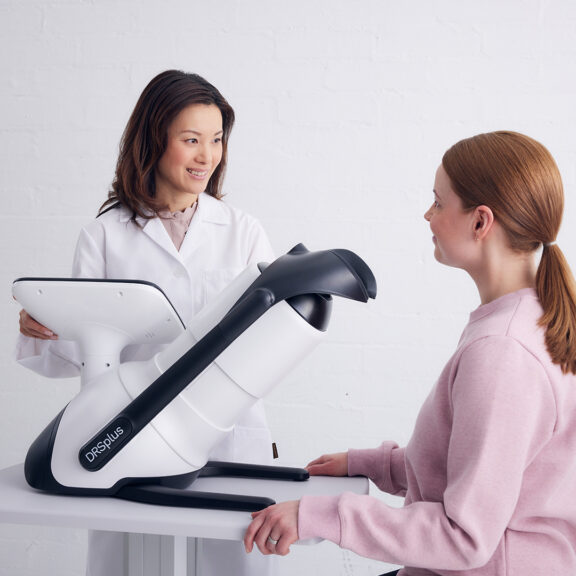
iCare DRSplus fundus imaging system with TrueColor Confocal Technology
iCare DRSplus confocal fundus imaging system uses white LED illumination to offer high-quality TrueColor images. TrueColor Confocal Technology, which is considered a standard of high image quality, provides detail-rich images with greater image sharpness, optical resolution and contrast when compared to traditional fundus camera imaging.
The fast and fully automated iCare DRSplus permits imaging through pupils as small as 2.5 mm, without need of dilation, ensuring a comfortable patient experience. This easy-to-use device offers the advantage of quick examination time and helps speed up workflow at clinics.
Key features
TrueColor Confocal Technology
Multiple imaging modalities including Red-free, Infrared, external eye and stereo view imaging
2.5 mm minimum pupil size
Fast, easy and fully automated operations
Mosaic function which creates retinal panoramic views up to 80°
Remote Viewer that allows for reviewing from devices on the same local area network
Remote Exam feature enables executing an exam from a distance
Different imaging modalities for detail-rich imaging
TrueColor Confocal Technology with white LED, promotes detailed 45° retinal images and allows to scan through cataract to aid clinicians in the diagnosis and documentation of ocular disease.
High resolution red-free filtering enhances visualization of retina vasculature, blue images provide improved view of the Nerve Fiber Layer (RNFL) and red channel allows light to penetrate into the deep layers of the retina. Infrared light provides detailed information corresponding to the choroid. External eye imaging can document eye surface and cornea conditions. Stereo viewer technology creates improved 3D perception of the disc. The mosaic function option automatically combines different retinal fields without user intervention, creating panoramic views up to 80°.
TrueColor Confocal Technology with white LED gives detailed 45° retinal images
TrueColor Confocal Technology with white LED, promotes detailed 45° retinal images and allows to scan through cataract to aid clinicians in the diagnosis and documentation of ocular disease.
Patient-friendly and comfortable device
iCare DRSplus Confocal Technology speeds up exam time while ensuring a comfortable patient experience. The device permits imaging through pupils as small as 2.5 mm and this non-mydriatic capability eliminates need for dilating drops and dark environments. With reduced flash intensity for softer effect on the pupil and quick and easy patient positioning, iCare DRSplus is a patient-friendly device.
Fast, efficient, and fully computerized operations
iCare DRSplus is an easy-to-use and intuitive imaging system that requires minimal staff training to obtain the highest quality images. iCare DRSplus offers live IR preview which contributes to an efficient capture workflow, while the filtering option makes image post-processing simpler. The fully-automated capabilities include auto-alignment, auto-focus, auto-exposure, auto-capture and auto-montage (optional). Quick and easy patient positioning and speedy examination saves time and makes the workflow efficient.
Image reviewing made easy
iCare DRSplus capacitive touch screen allows for easy magnification and review of images’ details. Additionally, the optional remote viewer feature makes remote data review effortless, allowing any computer or laptop on the same local area network (LAN) to review iCare DRSplus images remotely, enabling data access and detailed analysis on multiple review stations.
Remote Exam enables social distancing
In the fight against COVID-19, healthcare professionals are required to apply special social distancing precautions to minimize the spread of the virus. We are working closely with healthcare professionals during this difficult period to provide appropriate support and tools to facilitate social distancing during patient exams.
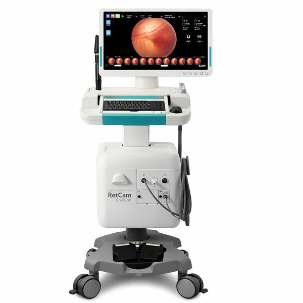
RETCAM Envision Ophthalmic Imaging System is a high-performance widefield retinal imaging platform designed for pediatric, neonatal, and adult retinal examinations. Widely used for ROP screening, retinal pathology documentation, and teaching, RETCAM Envision is supplied globally by @ErnestVision, a trusted ophthalmic equipment exporter from India serving hospitals, eye institutes, and specialty clinics.
RETCAM Envision Ophthalmic Imaging System Supplier in Washington, D.C.
RETCAM Envision Ophthalmic Imaging System Supplier in London, UK
RETCAM Envision Ophthalmic Imaging System Supplier in Riyadh, Saudi Arabia
RETCAM Envision Ophthalmic Imaging System Supplier in Bangkok, Thailand
RETCAM Envision Ophthalmic Imaging System Supplier in Canberra, Australia
RETCAM Envision Ophthalmic Imaging System Supplier in Singapore
RETCAM Envision Ophthalmic Imaging System Supplier in Kingston, Jamaica
🔬 Product Overview – RETCAM Envision Ophthalmic Imaging System
System Type: Widefield Retinal Imaging System
Applications: ROP screening, pediatric ophthalmology, retinal disease documentation
Users: Hospitals, NICUs, eye institutes, teaching centers
Imaging: High-resolution digital retinal capture
Design: Mobile, clinic-friendly, easy data storage & review
Supply: New system with export-ready support
⚠️ For professional ophthalmic and pediatric clinical use only.
📦 Why Choose @ErnestVision
Specialized ophthalmic diagnostic equipment exporter from India
Trusted by hospitals, NICUs & eye institutes worldwide
Complete technical documentation & export assistance
Global shipping via DHL, FedEx, Aramex
Dedicated pre-sales & after-sales coordination
📞 Contact – ErnestVision
@ErnestVision | @ErnestImpex
🌐 www.ernestvision.com | www.ernestimpex.com
📧 exports@ernestpharmaceuticals.com
📲 WhatsApp: +91 93599 02383
🔗 https://wa.me/919359902383
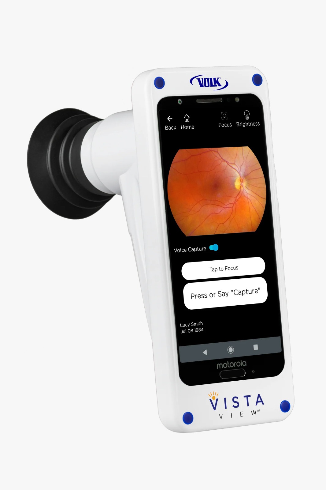
The Volk Fundus Camera VistaView is a portable, handheld, mydriatic retinal camera that uses a smartphone for its interface and workflow. It is designed to be affordable and easy to use, allowing eye care professionals to capture high-quality, 55° retinal images anywhere, such as in waiting rooms, nursing homes, or during mobile screening campaigns. Key features include on-device patient data management, instant report generation, and image export capabilities.
Key features and description
Portability: Its lightweight and ergonomic design makes it easy to carry and use in a variety of settings, including patient homes or nursing homes.
Integrated workflow: It connects to a smartphone to manage patient data, take images, review findings, add notes, and generate reports directly on the device.
Image quality: Powered by Volk optics, it captures sharp, 55° fundus images with user-adjustable illumination for patient comfort.
Ease of use: The device is designed for simplicity, with features like voice command capture, making it easy for any staff member to use, including residents.
Advanced software: It includes an interactive touchscreen for detailed review, a red-free filter to identify lesions, and the ability to create password-protected reports for sharing with other professionals.
Affordability: It aims to make high-quality retinal imaging accessible and affordable, eliminating the need for heavy financing associated with traditional, larger fundus cameras.
Patient-friendly: User-adjustable illumination and a compact design allow for more comfortable exams, especially for photophobic or less mobile patients.
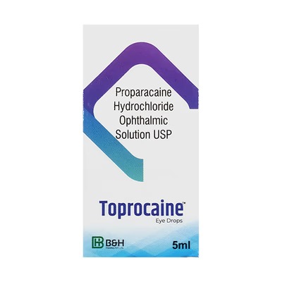
Composition:
Each ml contains Proparacaine Hydrochloride 0.5% w/v
Description:
TOPROCAINE Eye Drops 5ml is a local anesthetic ophthalmic solution designed to provide rapid and temporary relief from ocular pain and discomfort during minor eye procedures, diagnostic tests, or irritation caused by foreign particles. The active ingredient, Proparacaine Hydrochloride, ensures fast-acting numbing effect with minimal systemic absorption.
Its gentle formulation allows for comfortable application, making it suitable for both adults and children under medical supervision.
Uses & Benefits:
Provides local anesthesia for minor eye procedures
Relieves ocular discomfort, irritation, or foreign body sensation
Ensures rapid onset of action
Supports accurate ophthalmic examinations and procedures
Safe for short-term use under professional supervision
How It Works:
Proparacaine Hydrochloride blocks nerve signal transmission in the cornea and conjunctiva, temporarily numbing the eye surface and preventing pain sensation.
Directions for Use:
Instill one drop in the affected eye(s) as advised by your ophthalmologist
Avoid contact between the dropper tip and any surface
Use only as needed for short-term relief
Storage Instructions:
Store in a cool, dry place below 30°C, away from sunlight. Discard any remaining solution four weeks after opening.
Pack Size:
5ml sterile ophthalmic solution bottle
Composition:
Each ml contains Proparacaine Hydrochloride 0.5% w/v
Description:
Aurocaine Eye Drops 5ml is a local anesthetic ophthalmic solution designed to provide rapid and temporary relief from ocular pain and discomfort during minor eye procedures, diagnostic tests, or irritation caused by foreign particles. The active ingredient, Proparacaine Hydrochloride, ensures fast-acting numbing effect with minimal systemic absorption.
Its gentle formulation allows for comfortable application, making it suitable for both adults and children under medical supervision.
Uses & Benefits:
Provides local anesthesia for minor eye procedures
Relieves ocular discomfort, irritation, or foreign body sensation
Ensures rapid onset of action
Safe for short-term use under professional supervision
Supports accurate ophthalmic examinations and procedures
How It Works:
Proparacaine Hydrochloride blocks nerve signal transmission in the cornea and conjunctiva, temporarily numbing the eye surface and preventing pain sensation.
Directions for Use:
Instill one drop in the affected eye(s) as advised by your ophthalmologist
Avoid contact between the dropper tip and any surface
Use only as needed for short-term relief
Storage Instructions:
Store in a cool, dry place below 30°C, away from sunlight. Discard any remaining solution four weeks after opening.
Pack Size:
5ml sterile ophthalmic solution bottle.
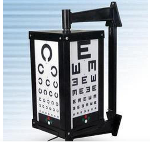
Eye Testing Drum Specification
Color
Black & White
Material
plastic
Size
6 Meter
Weight
Approx 1-2 Kg Kilograms (kg)
Length
6 Meter Millimeter (mm)
Eye Testing Drum Trade Information
Minimum Order Quantity
1 Piece
Supply Ability
2 Pieces Per Day
Delivery Time
7 Days
Main Export Market(s)
Western Europe, Australia, North America, Eastern Europe, Africa, Central America, Middle East, South America, Asia
Main Domestic Market
All India
About Eye Testing Drum
Eye Testing Drum which has a metal body and is strong and sturdy in nature. Our range of drums is internally printed as per snellen chart and is available with various tests namely duochrome test, friend test, spot light test and worth four dots test. These are remote controlled from a distance of 6 meter with the help of automatic switch.The Eye Testing Drum is a valuable tool that enables fast and accurate visual acuity assessments. Ideal for both medical and home uses, this device is simple to use and helps to accurately monitor the progression of a patient's vision. Our Eye Testing Drum offers a dependable method of reading and measuring eye health, making it a must-have tool in any vision-testing facility.
R-478 LED X-Ray View Box: High-Luminance Slimline Film Viewer – Global Export by Ernest Vision
In the precision-driven diagnostic landscape of 2026, the clarity of film interpretation remains a cornerstone of patient care. The R-478 LED X-Ray View Box is a professional-grade medical imaging accessory designed for the crystal-clear viewing of X-ray, CT, and MRI films. Utilizing advanced Surface Mounted Device (SMD) LED technology, this view box provides a uniform, flicker-free light source with a brightness of over 10,000 Lux, ensuring that even the most subtle clinical details are visible to radiologists and surgeons.
Ernest Vision, a specialized division of Ernest Pharmaceutical Pvt. Ltd., is a leading Exporter, Supplier, and OEM Partner from India. We facilitate the strategic distribution of the R-478 to Dubai, Muscat, Abuja, Yaoundé, and Jakarta, providing elite-tier, energy-efficient, and ultra-slim diagnostic tools for the world’s most advanced healthcare networks.
Technical Specifications: R-478 LED X-Ray View Box
The R-478 is meticulously built to provide maximum diagnostic accuracy while minimizing eye fatigue for medical professionals.
Product Features & Advanced Optics
Ultra-Slim Profile: Featuring a modern, space-saving design with a thickness of only 25mm to 30mm, making it ideal for both wall-mounting and desk-top use in crowded clinics.
High-Intensity LED Array: Equipped with long-life LEDs (rated for 100,000 hours) that provide a color temperature of 8000K to 9000K, perfectly mimicking natural daylight for optimal film contrast.
Uniform Luminance: Advanced light-guide plate technology ensures a luminance uniformity of >90%, eliminating “dark spots“ or glare that can lead to diagnostic errors.
Flicker-Free Performance: Digital high-frequency ballast eliminates the 50Hz/60Hz flicker associated with traditional fluorescent viewers, significantly reducing eye strain during long shifts.
Film-Sensing Auto-Switch: Features an intelligent Auto-Switch Sensor that illuminates the screen only when a film is inserted, drastically reducing power consumption and heat output.
Rotary Dimmer Control: Integrated step-less brightness adjustment allows the operator to fine-tune light intensity based on the density of the film (MRI vs. standard X-ray).
Performance Standards
Light Intensity: High-brightness output of 10,000 to 15,000 Lux.
Configurations: Available in Single, Double, Triple, and Quadruple (Four-Film) panel versions.
Viewing Area: Standard 14“ x 17“ per panel, compatible with all international film sizes.
Power: Multi-voltage support (AC 100-240V) for seamless integration into global hospital grids.
Strategic Advantages for 2026 Healthcare Facilities
Diagnostic Excellence: Essential for orthopedic and oncology centers in Dubai and Muscat, where high-contrast viewing is critical for pre-surgical planning.
Tropicalized Durability: The specialized acrylic viewing screen and aluminum-alloy frame are resistant to yellowing and warping in high-humidity climates like Jakarta and Cameroon.
Operational ROI: With a lifespan of over 10 years and 80% lower energy consumption than fluorescent models, it is the most cost-effective choice for government health tenders in Abuja and Yaoundé.
Infection Control: The seamless, flush-mount design is easy to sanitize with standard 2026 medical-grade disinfectant wipes.
Global Distribution: UAE, Oman, Nigeria, Cameroon, & Indonesia
Ernest Vision manages the end-to-end regulatory and logistical requirements to supply high-quality Indian diagnostic equipment to key international markets:
Dubai, UAE & Muscat, Oman: Supplying premium private radiology suites and government medical cities with Tier-1 LED film viewers.
Abuja, Nigeria & Yaoundé, Cameroon: Delivering durable, high-efficiency X-ray equipment for national healthcare modernization and teaching hospitals.
Jakarta, Indonesia: Supporting national healthcare expansion with standardized, high-tech diagnostic tools for regional hospital networks.
📞 Contact Ernest Vision — Medical Device & Ophthalmic Exporter
(Group of Ernest Pharmaceutical Pvt. Ltd.)
Partner with a verified leader for the bulk procurement of LED X-Ray Viewers and Diagnostic Imaging Accessories.
🌐 Websites: www.ernestvision.com | www.ernestimpex.com | www.oncologymedicinesupplier.com
📧 Email: exports@ernestpharmaceuticals.com
📦 Business Type: Exporter | Supplier | OEM Partner
📲 WhatsApp: +91 93599 02383
🔗 Instant Chat: https://wa.me/919359902383
HOSPITAL FURNITURE
Still searching for hyaluronidase uses in imaging tests?
 Chat with us