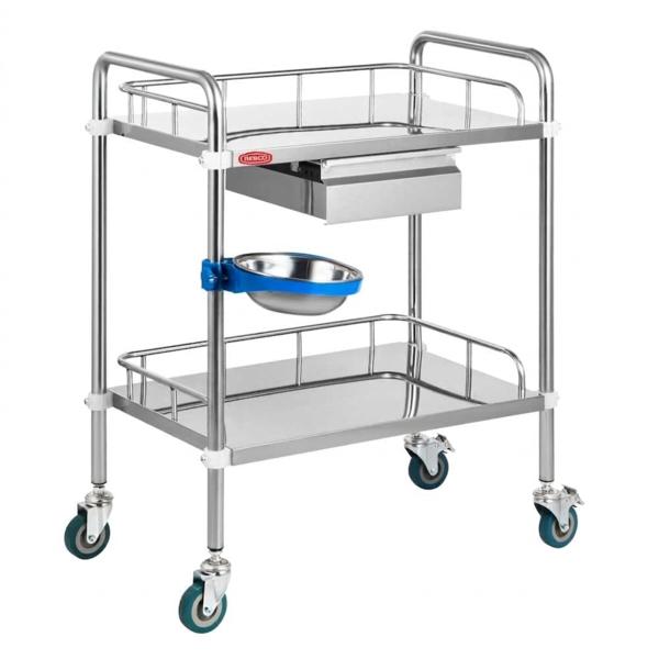
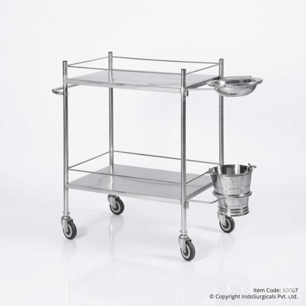
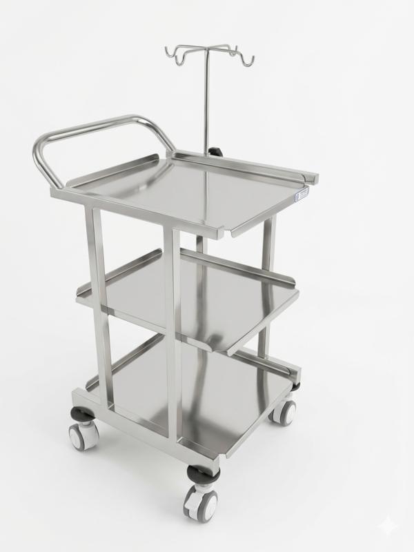
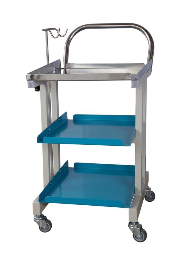
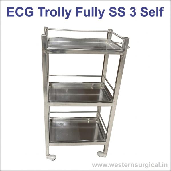
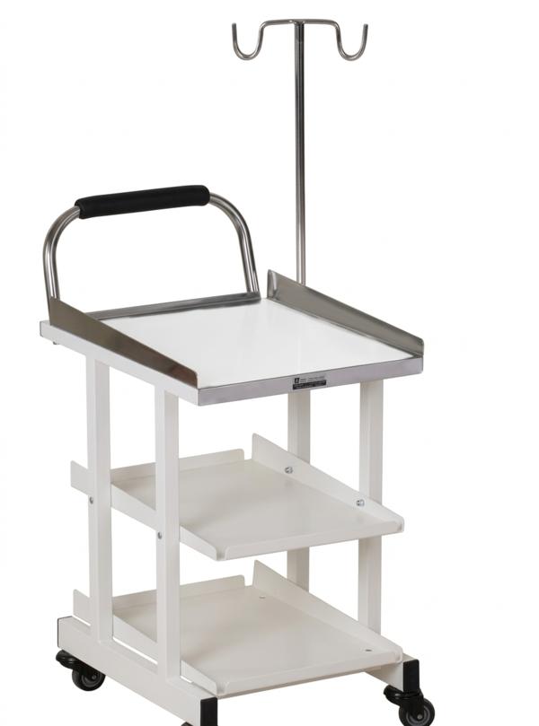
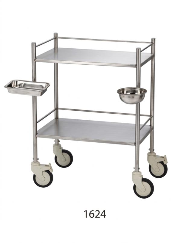
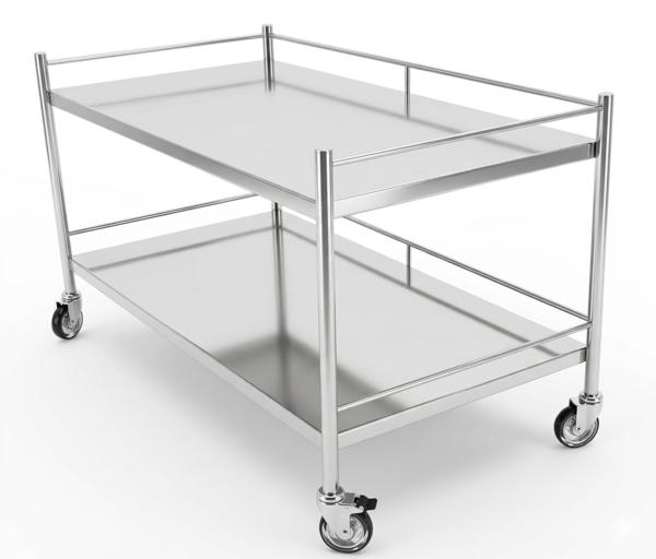
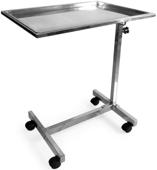
Dubai
+919359902383
+919359902383

Products

6814772a5d263414004dd4a4 Card 2

product image
Ophthalmic Devices

Enhanced magnification for advanced procedures If you can’t see it, you can’t work on it, which is why Keeler Prismatic loupes offer magnifications of 3.5x, 4.5x and 5.5x in 3 working distances. These loupes are higher magnification with prismatic optics, guaranteeing edge to edge clarity and precise colour rendition. Keeler Prismatic loupes are laser aligned and treated with anti reflective coating to produce a superior image. Features: Higher magnification. Laser aligned optics. 3 Working distances. Anti reflective coating. Available with Keeler frame and K-LED. Available on headband. Superb quality. Lightweight. SKU: Prismatic loupes 4.5x magnification Contents: Keeler lightweight loupes Pair of protective caps Flip lever. Instructions CD (Optional) Keeler sport frame. Lightweight protective shield. 3 way adjustable hinge. 2 x adjustable nose pieces. Plano prescription insert. Neck cord. Lens cloth. Protective case.

6814772a5d263414004dd4a4 Card 2

product image
Ophthalmic Devices

Power 1 kW to 3 kW Brand EYE PLUS Categories optical equipment Country of Origin Made in India Availability In Stock Lens Groover Machine, designed for precision grooving of semi-rimless and rimless eyeglass lenses. This is based on which are widely used in optical workshops and clinics. 🌀 Lens Groover Machine – Full Specifications⚙️ Technical Features Feature Specification Groove Position Adjustable: Front, Center, Rear Groove Depth 0 to 0.7 mm Groove Width 0.65 mm Lens Thickness Range 1.5 mm to 11 mm Lens Diameter Range 18 mm to 70 mm Grooving Types Central groove, Front arc groove, Rear arc groove Material Compatibility PC (polycarbonate), CR-39 plastic, High-index plastic, Glass, Trivex 🛠️ Mechanical & Build Body Material: Stainless steel panel with grooving slot Diamond-coated grooving wheel (imported) Drive System: DC motor with stable torque Manual feed or auto-feed depending on model Control: Manual dial or lever for groove depth and position Adjustable lens holder with anti-slip grip 🔌 Power & Electrical Feature Specification Voltage 220V / 50Hz or 110V / 60Hz (region dependent) Power Consumption 80W Motor Type DC motor (low noise, high precision) 📐 Dimensions & Weight Feature Specification Machine Size 295 mm (L) × 250 mm (W) × 190 mm (H) Net Weight ~3 kg Package Dimensions Approx. 320 mm × 270 mm × 220 mm 🧰 Included Accessories Diamond grooving wheel (pre-installed) Lens holder clamps Power cord User manual Optional: Dust cover or cleaning brush 🛡️ Safety & Compliance CE-certified components (varies by manufacturer) Overload protection circuit Non-slip base for stable operation

6814772a5d263414004dd4a4 Card 2

product image
Ophthalmic Devices

Pocket Ophthalmoscope The ophthalmoscope with superior aspherical optics Features Lightweight and compact. Halogen illumination provides brighter, whiter, longer lasting light. Sealed optice keep instrument clean and dust free . Aperture dial offers six selections Glass condensing lens. Includes red-free and cobalt blue filter. Standard AA Batteries with interchangeable heads for otoscope and ophthalmoscope Handle artwork customization available (minimum order 5000 units) Ordering Information 12850 Pocket Junior Ophthalmoscope complete with AA handle and batteries 12851 Pocket Junior Ophthalmoscope complete with AA handle, batteries and case 03900 Replacement Lamp

6814772a5d263414004dd4a4 Card 2

product image
Ophthalmic Devices

Cutting through, for a clear occular view The B-scan probe is designed to provide you with crystal clear images of the posterior segment of the eye, whilst also visualising possible occular tumors, retinal detachments, vitreous hemorhages and cataracts, for a prompt and reliable diagnosis. With built-in tools and automatic functions to optimise your workflow, the B-scan probe is a reliable partner for your ultrasound examinations, allowing you to spend more time with your patients without interruptions. Working in harmony with Keeler Connect software (which can be plugged into any Windows™ laptop or desktop computer), B-Scan Plus Connect is intuitively designed and fully portable, making it the perfect companion for busy eye imaging professionals, either in clinic or on-the-go The B-Scan probe is also fully compatible with the Keeler 4Sight™ ultrasound system – our all-in-one ultrasound solution. Benefits: Exceptional optics with 0.015mm image resolution High image resolution Smooth zoom technology – getting you two times closer without distortion Device portability Extensive scanning – dual frequency (12 or 15MHz) with a 60deg sweep angle, for comprehensive image or video capture Competitive warranty and great customer support Fully compatible with Keeler 4Sight™ – giving you unmatched storage space State of the art – software includes 12 measuring callipers, scan labelling tools, screen-shot capture, auto-print and reporting functions for data export Accessories included: Software installation, USB stick, probe holder, footswitch, wireless mouse, and ophthalmic gel. High resolution accuracy and reliability Get ready to enhance your examinations, with a reliable diagnosis, prompt referral, increased revenue and an enhanced patient experience! Record and save unlimited images and videos with the B-scan probe and its proprietary software, 4Sight™, to assist with diagnosing, record keeping and even teaching future generations of eye care professionals. Smooth Zoom The B-scan probe utilises ‘Smooth Zoom’ technology to give you 2x full image zoom without distortion, to give you the extensive detail you need when performing ultrasound examinations. Device portability For the ultimate portability and flexible usage, the B-scan probe can be plugged into any Windows™ laptop or desktop computer, making it the perfect companion for busy eye care professionals on the go. 4Sight compatibility for unmatched storage space Record and save unlimited images and videos with the B-scan probe and its proprietary software, 4Sight™, to assist with diagnosing, record keeping and even teaching future generations of eye care professionals. Versatile usage As well as imaging the internal structure of the eye, the B-scan probe works to visualise possible tumours, haemorrhages, detachment and diseases, in order to assist with future surgery and provide the best results possible. Extensive scanning The B-scan probe works on a dual frequency (12 or 15 MHz) and compiles a series of 256 A-scans over a 60º sweep angle, providing you with a comprehensive range of images and videos. State of the art features Conveniently included in the B-scan software are 12 measuring callipers, scan labelling tools, screen-shot capture, auto-print and reporting functions for data export, to ensure you have everything you need all in one place. Features – Keeler Connect software: Intuitive interface Industry leading portability Ultrasound compatibility Great value Competitive warranty and great customer support Features – Keeler B-Scan probe: 0.015mm image resolution is among the highest in the medical industry. Improved probe providing sharper, more focused images due to the elimination of signal loss. Adjustable film loops that range from 2 seconds to 17 seconds. Unlimited adjustable and transferable video recording. Fully adjustable gain-control before, during or after scan. Easy to set up defaults, smooth customer interface and automatic EMR exports to decrease examination time, increase patient throughput, and increase profit. Fully upgradable software. EMR compatible. DICOM ready. Multiple measuring velocities for ACD/VCD. PDF reporting. Archive data on a network or external location. Specification: Weight: 6 oz. Dimensions: 7” long X 1.25” diameter Frequency: 12 or 15 MHz Axial resolution: 0.015 mm Electronic lateral resolution: 0.085 mm Electronic gain: 0-112 dB Adjustable gamma: Linear, S-Curve, Log, Color Scanning Angle: 60° Field of view: 12 MHz = 64 mm or 15 MHz = 52 mm Frame rate per second: 15, 30 max Sampling Rate: 2048 (points per line) Vectors per frame: 256 Focal point: 22 mm Focal zone: 20 mm TGC: Yes Frozen image gain adjustment: Yes Zoom: 2x optical zoom, 8x Maximum Reports: PDF reporting Snapshot format: jpeg, bmp, png, tiff, gif Data archive/ Export capability: Yes

6814772a5d263414004dd4a4 Card 2

product image
Ophthalmic Devices

High quality visuals and accurate biometric measurements, on demand This unit contains everything you need to get you up and running on Keeler 4Sight™ – our all-in-one ultrasound solution. The most demanding cataract surgery requires powerful equipment and the A-Scan is the single most effective solution for measuring, calculating and refining your refractive cataract surgical outcomes. Designed to take accurate biometric measurements, the A-Scan probe can be used to measure ocular axial length in contact or immersion modes and it is particularly useful for patients or animals with fixation difficulties or dense cataracts. Compatible with both Keeler Connect software (which can be used on any laptop or desktop computer) and Keeler 4Sight™ ultrasound system, the A-Scan probe is intuitively designed and fully portable, making it the perfect companion for busy eye imaging professionals, either in clinic or on-the-go. Benefits: Improved visuals Easy to use interface with time-saving functionality Wavelength spike patterns for accurate and precise measurements Includes six IOL formulas Includes four post-refractive formulas Competitive warranty and great customer support Fully compatible with Keeler 4Sight™ – giving you unmatched storage space High quality visuals Record and save unlimited images and videos with the B-scan probe and the Keeler 4Sight™ software, to assist with diagnosing, record keeping and even teaching future generations of eye care professionals. Featuring optimised lens constants for improved visuals, the A-scan probe allows you to see visuals that would otherwise be obscured by cataracts or vitreous haemorrhages, providing superior surgical outcomes for your patients. Easy to use interface The smooth interface makes the A-scan probe easy to use, and allows you to create default settings that you can go back to again and again without interruptions. The system also automatically creates electronic medical records for your patients, giving you peace of mind for future examinations. Wavelength spike patterns Working on 10 MHz broadband, the A-Scan probe can be used to measure ocular axial length in both contact or immersion modes. Each measurement with the A-scan captures up to 5 wavelength spike patterns, using independent velocities for each portion of the eye, as well as the average and standard deviation of each anatomical measurement to give you the most accurate results. IOL formulas Includes 6 IOL formulas: Hoffer Q, SRK/T, Holladay I, Haigis, SRK II, Binkhorst II. Post refractive formulas The A-scan also has post refractive measurement capabilities, using 4 formulae: Contact Lens, Entered, Clinical History and Shammas Clinical. Great value At Keeler, we believe that value is more than the price alone. We look at every detail and quality components, results and high build quality shouldn’t cost the earth. Support when you need it True partnerships don’t stop, our industry-leading warranty and service programs, as well as our live help facility offer dependable support for the life of the product. Features – A-Scan probe: Immersion and contact modes Customizable lens, ACD and VCD materials for new emerging implant/chamber materials Optimized lens constants for superior surgical outcomes Easy to set up defaults, smooth customer interface and automatic EMR exports to decrease examination time, increase patient throughput, and increase profit Automatic alignment detection and sclera recognition help eliminate marginally aligned scans Seamless work flow integration with IOLMaster 500 from ZEISS Fully upgradeable software Unlimited data storage based on PC/network storage EMR capability with automatic transfer of data DICOM ready Unlimited IOL Lens groups Piggyback calculation ability for ICL implant patients Rx surgery calculations for an aging Lasik population PC based platform PDF reporting Specification: Capture Modes: Automatic or Manual Clinical Accuracy: 0.1mm Custom Eye Type Settings: Unlimited with independent ACD, Lens, VCD velocities. Data Export: EMR/EHR compatible, Network ready Default Eye Type Settings: Phakic, Dense Cataract, Silicone Filled, Pseudo PMMA, Pseudo Silicone, Pseudo Acrylic, Aphakic Dimensions: 5.4″W x 3.7″L x 2.5″H Electronic Resolution: 0.016mm Frequency: 10MHz Gain: 30-100dB IOL Data Storage: Unlimited Measurement Range: 0.01-63.6mm Measurements: ACD, Lens thickness, VCD, Axial Length Methods: Immersion or contact Patient Data Storage: Unlimited Post Refractive Formula Methods: Clinical History, Contact Lens, Shammas Clinical Printer: Any supported by PC Weight: 15.1 oz

6814772a5d263414004dd4a4 Card 2

product image
Ophthalmic Devices

Single-use iCare probes ensure patients’ safety The probes are designed for single use only, and they should not be cleaned or re-used. Multiple-use of the probe can cause cross-contamination. Cleaning or mishandling the probe can bring damage and shorten the lifetime of the device as well as affect the measurement result. To ensure patient safety and the accuracy of IOP measurements, we recommend using the original iCare probes only. Repeatable IOP measurements The probe consists of gold-plated metal wire with a small plastic tip made of medical-grade plastic. During the measurement procedure, the probe makes brief, momentary contact with the eye’s cornea six consecutive times for each eye. The patient barely notices the contact. The probe’s weight and straightness tolerances are very tight to ensure repeatable IOP measurements. Hygienic and safe – no contamination risk Owing to rebound technology, use of an iCare tonometer does not create microaerosol formations, because measuring requires no disinfection, anesthesia, drops, air, or other preparation. Easy to insert and remove The iCare probes are very easy to insert in and remove from the probe base. The probes are supplied in individual protective probe tubes. After removing the probe from the tube, it is only needed to allow the probe to slide down to the probe base. The tonometer automatically magnetizes and holds the probe in place.

6814772a5d263414004dd4a4 Card 2

product image
Ophthalmic Devices

Usage/Application ENT Surgery Material Stainless Steel Categories Holding Instruments Material Grade Surgical Grade Surface Finish Gloss Brand Max Plus Country of Origin Made in India Availability In Stock The Max Plus Epilation Forceps are a specialized medical instrument designed for the precise and hygienic removal of individual hairs or foreign bodies. Unlike standard tweezers, these forceps are meticulously crafted with extremely fine, pointed tips that allow for the secure grasping of a hair follicle without damaging the surrounding skin. This level of precision is critical for delicate procedures, particularly in ophthalmology and dermatology. Key Product Features: Fine, Pointed Tips: The tips are precisely aligned and sharpened to grasp a single hair follicle firmly, preventing breakage and ensuring complete removal. Ergonomic Design: The forceps are designed for optimal balance and a comfortable grip, minimizing hand fatigue and allowing for greater control and accuracy. Premium Surgical Stainless Steel: Made from high-quality, medical-grade stainless steel, the forceps are completely resistant to corrosion, rust, and are built to withstand the extreme temperatures of repeated sterilization cycles. Versatile Use: While primarily used for epilation, their fine tips make them suitable for a variety of tasks requiring precision, such as handling small dressings or retrieving minute foreign bodies. Fully Autoclavable: The high-quality stainless steel material allows for complete and effective steam sterilization, ensuring the highest level of infection control. Uses of the Product The Epilation Forceps are a vital tool with numerous critical applications in a clinical setting. Their primary uses are: Ophthalmology: They are a standard tool for the removal of misdirected or ingrown eyelashes, a condition known as trichiasis, which can cause significant eye irritation and damage. Dermatology: They are used by dermatologists and estheticians for the removal of stubborn, coarse hairs, particularly those causing ingrown hairs or cosmetic concerns. Pre-operative Preparation: They can be used to remove individual hairs from a surgical site to ensure a completely smooth surface for incision, reducing the risk of infection. Foreign Body Retrieval: The fine tips allow for the careful grasping and removal of small, superficial foreign bodies from the skin. How to Use the Product Sterilization: Before each use, the forceps must be thoroughly cleaned and sterilized, typically through autoclaving, to prevent cross-contamination. Handling: The sterilized forceps are carefully removed from their sterile pack. The fine tips are always kept in view to prevent accidental injury or damage. Application: The tips are used to carefully grasp the hair near the follicle, and a swift, gentle pull is applied in the direction of the hair growth. Cleaning: After the procedure, the forceps are immediately taken for cleaning and re-sterilization. The tips should be thoroughly cleaned to remove any debris before autoclaving. Product Categories According to Surgical Procedures Ophthalmology Instruments: Their use in trichiasis and eye care makes this a core category. Dermatology Instruments: They are essential tools for dermatological procedures. Minor Surgery Instruments: They are commonly used for simple, non-invasive procedures. Reusable & Autoclavable Tools: As a reusable and autoclavable instrument, it is a key part of a clinic's sterile toolkit.

6814772a5d263414004dd4a4 Card 2

product image
Ophthalmic Devices

With the aid of our proficient professionals, we have been able to offer qualitative Viscoelastic Gel. The products offered by us are made by a team of educated professionals, keeping the industry laid norms and guidelines in mind. All our products are widely appreciated by the respectable clients for having features like accurate composition, safe usage, precise pH value and longer shelf life. Some of the specifications of offered gel is given below: Composition: Hydroxypropyl Methyl cellulose I.P. 2% w/v (High Viscosity) Sterile Isotonic Base q.s. Packaging:5 ml vial, 2 ml pre-filled syringe with cannula

6814772a5d263414004dd4a4 Card 2

product image
Ophthalmic Devices

CASTROVIEJO SUTURE TYING FORCEPS MOSQUITO HEMOSTAT FORCEPS CURVED MOSQUITO HEMOSTAT FORCEPS STRAIGHT KELMAN MCPHERSON FORCEPS ANGLED UTRATA CAPSULORRHEXIS DELICATE FORCEPS IRIS EYE DRESSING FORCEPS IRIS EYE TISSUE FORCEPS HARMS COLIBRI DELICATE FORCEPS, 1X2 TEETH WITH TYING PLATFORM BARRAQUER WIRE EYE SPECULUM BARRAQUER MICRO NEEDLE HOLDER CURVED BARRAQUER MICRO NEEDLE HOLDER STRAIGHT CASTROVIEJO CALIPER KNAPP LACRIMAL RETRACTOR 4 PRONGS DESMARRES LID RETRACTOR , SIZE 0 DESMARRES LID RETRACTOR , SIZE 3 SIEGEL ROUND SCALPEL BLADE HANDLE #3 DESMARRES CHALAZION FORCEPS , LARGE ADSON MICRO TISSUE, 1X2 TEETH FORCEPS ADSON MICRO DRESSING SERRATED FORCEPS KNAPP EYE SPECULUM MCPHERSON VANNAS SCISSORS CURVED CASTROVIEJO SCISSORS 4.5“ STRAIGHT CASTROVIEJO CORNEOSCLERAL SCISSORS RIGHT CASTROVIEJO CORNEOSCLERAL SCISSORS LEFT HOSKIN #19 TISSUE FORCEPS MICRO GROOVED TIPS STRAIGHT BISHOP HARMON MICRO TISSUE, 1X2 TEETH FORCEPS CASTROVIEJO NEEDLE HOLDER STRAIGHT 5.5“ CASTROVIEJO NEEDLE HOLDER CURVED 5.5“ JONES TOWEL CLAMP EYE IRIS SCISSORS 4.5“ STRAIGHT

6814772a5d263414004dd4a4 Card 2

product image
Ophthalmic Devices

Shinnipon Japan to Labomed Max features are computer designed, multi-coated optics that provide unsurpassed viewing of ocular structures, the operator's field of view is maximized for precise visualization of ocular defects at the periphery. Specifications Binocular Microscope Eye Pieces : 10x, 15x Objective : 1x, 1.6x Total Magnification : 10x, 16x and 24x Illumination Unit Slit Image Rotation : 0 to 180° Tilting Illumination : Up to 20° Filter Disc : Cobalt Filter, Green Filter, Yellow Filter Natural Density and Open Aperture Slit Disphragm Disc : Wedge Shaped Diaphragm Of Infinitely Variable Slit Lengths Halogen Lamp : 6 Volt 20 Watts tandard Accessories 15x Eyepieces Plastic Breath Shield Replacing Mirror Hruby Lens Dust Cover and Fuse Testing Rod Replacing Lamp one piece for illumination unit Optional Accessories Power Table / Mechanical Table Optional accessories are listed as separate items in “Accessories“ section under products, there brought separately.

6814772a5d263414004dd4a4 Card 2

product image
Ophthalmic Devices

Magnification 3.5x Working Distance 34 cm Application Hospital Field of View 3.5x:- 5.5 cm Brand Keeler Model Number Prismatic Availability In Stock Keeler Prismatic loupes offer magnifications of 3.5 working distances. These loupes are higher magnification with prismatic optics, guaranteeing edge to edge clarity and precise colour rendition. Keeler Prismatic loupes are laser aligned and treated with anti reflective coating to produce a superior image. Contents: Keeler prismatic loupes Pair of protective caps Flip lever Instructions CD (Optional) Keeler sport frame Lightweight protective shield 3 way adjustable hinge 2 x adjustable nose pieces Plano prescription insert Neck cord Lens cloth Protective case Features:- Superb Quality Available with Keeler Frames and K-LED Available on Headband High Magnification Laser Aligned Optics 3 Working Distances Anti Reflective Coating

Phone Number

Mon-Thu: 10 AM - 2 PM • Fri: 3 PM - 7AM

Other Website Visit our other website
Address

Dubai - United Arab Emirates, Dubai