Dubai, United Arab Emirates
+919359902383
+919359902383

Items tagged with 'automated endothelial cell analysis'

6814772a5d263414004dd4a4 Card 2

product image
Ophthalmic Devices

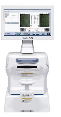
The All-New, Blazing Fast, Fully-Automated Specular Microscope > Widescreen Touch Panel PC mounted > Fully automated, integrated database > Location specific data samples > Robust Analysis Methods CellChek®️️️ 20 & 20 PLUS The All-New, Blazing Fast, Fully-Automated Specular Microscopes Process bilateral exams with one touch in about 40 seconds Konan specular microscopes were recognized as the gold standard specular microscopes in the minutes from an FDA panel meeting* The Gold Standard, Redefined. Faster, Easier Endothelial Imaging CellChek®️️️ 20 is Konan Medical’s new, non-contact specular microscope that can capture and analyze bilateral exams with one touch in under 40 seconds. The new easy-to-use “Simple Mode” enables one-touch, fully-automated endothelial image capture with analysis, reporting, and exporting of data.  Clinical Applications Glaucoma, cataract, & refractive surgery. Corneal disease management. Contact/specialty contact lens fittings. Routine eye care N Clinical Benefits Visualize endothelial cells with 40x magnification compared to slit lamp bio-microscopy Identify pre-existing low density and dystrophies that may affect positive surgical outcomes Confirm recommended minimum cell density/morphology for scleral/specialty contact lenses  Regulatory FDA 510(k) Cleared | CPT Code 92286 CE Marked Health Canada Licensed Blazing Fast The new, easy-to-use “Simple Mode” enables one-touch, fully-automated endothelial image capture with analysis, reporting, and exporting of data. The completely reimagined CellChek®️️️ 20 is noticeably improved and simplified; remove the device from the box, connect to power, turn it on, and start imaging. Konan’s gold standard Center Method™ and Flex Center Method™ are now fully-automated. Automated Center Method displays cell center counted densities and morphometric indices of both eyes immediately after images are captured – all in under 40 seconds. Key New Features With CellChek®️️️ 20 you can capture, analyze and print/export a bilateral exam with just one touch. The new, easy-to-use “Simple Mode” makes running a bilateral exam faster and easier than ever before. CellChek®️️️ 20 reduces testing time significantly, allowing you to test patients quickly and efficiently.  One-touch imaging Fast, one-touch imaging of both eyes including positioning, alignment, imaging capture, analysis and print-out/exporting data.  Optimized touch functions Easy touch-friendly interface makes capturing, analysis, and reporting fast and efficient. Also features pinch-to-zoom image adjustment.  25% higher resolution Improved resolution allows better visualization of single cells and cell walls. Superb cell border tracing and guttae exclusion.  Auto-capture/retry This improved technology allows for successful imaging of difficult or challenging cases.  30% larger image area The field of view has been increased to 0.25mm x 0.55mm, enabling better corneal visualization and more cells to be analyzed. CellChek®️️️ 20 PLUS CellChek®️️️ 20 PLUS adds near-limbal imaging to the standard model. It extends the limits of corneal endothelial image capture adding locations to 4.5mm from center. Six additional fixation points, 13 total. Flexible Touch Screen The flexible 10.6” wide touch screen can be turned and tilted 180°, providing better ergonomics, space savings, and flexibility with technician use from any of the four sides: front, back, left, and right. Fully-Automated Analysis CellChek®️️️ 20 offers fully-automated: Center Method; Flex-Center Method; and Auto-Trace analysis. These analysis methods provide one-touch, high precision results FAST. These fully-automated analysis methods are offered alongside the traditional manual Center and Flex-Center analysis methods, offering a total of 6 analysis options. Konan’s Center Method is mentioned in FDA panel minutes as being the “gold standard”, and is used by virtually every professional reading center. New Auto Center Method With the new Auto Center Method, the center of each individual, visible cell is automatically marked. Guttae and other dark regions are automatically excluded. New Auto Flex-Center Method With the new Auto Center Method, the center of each individual, visible cell is automatically marked. Guttae and other dark regions are automatically excluded. Clinical Benefits Cataract Surgery and Premium IOLs Low endothelial cell counts and pre-existing dystrophies can markedly reduce the potential for positive surgical outcomes from an otherwise uneventful cataract surgery. Surgeons are finding these data points critical when recommending premium IOLs: verify and document pre-operatively that the cornea is not suspect to more likely post-op complications difficult to explain with the investment in premium IOLs.

6814772a5d263414004dd4a4 Card 2

product image
Ophthalmic Devices

CellChek® D | D+ Eye Bank Specular Microscopes Designed to improve patient outcomes, CellChek® D+ redefined specular microscopy in Eyebanking. It was quickly and widely adopted by many of the world’s finest Eye Banks. CellChek® D+ Introducing the first multi-imaging system for donor corneal analysis. CellChek® D+ provides an amazing view into the cornea that simply has never been seen before defining the structure of the endothelium, stroma and epithelium. Comprehensive imaging to assist both eye banks and corneal surgeons to make better decisions on implantable material. Now featured in Cornea June 2018 Supplement: Current and New Technologies in Corneal Donor Tissue Evaluation Comparative Image Atlas. Especially check out S7 “Wide-Field Ex Vivo Dual Imaging Microscopy.” Clinical Benefits Provides remarkable views of endothelium, intra-stomal structure and the epithelium For the first time, visualize blood cells, fungus, rough keratome cuts, dead cells, and epithelial anomalies New “Donor Enhanced” Imaging System CellChek® D+ features a patent applied for “Donor Enhance” imaging system that provides remarkable views of endothelium, intra-stomal structure, and the epithelium. The range of new information from which to make better clinical decisions is spectacular (pun intended): blood cells, fungus, rough keratome cuts, dead cells, epithelial anomalies. Never have we had this view of pre-implanted material. Corneal surgeons are finding this information to be illuminating in helping to understand graft success and failure. Broad overview but with large multi-sample analytic areas CellChek® D and D+ feature a new finder feature with digital measuring / documenting tools. Patented Cellular Analysis Methods All Konan specular microscopes feature the Center Method of analysis. Center Method is mentioned in FDA panel minutes as being the “gold standard” and is used by virtually every professional reading center for independent assessment of corneal endothelial analytics.¹ Unlike full-auto assessments, the Center Method provides high precision and repeatability for specular images in which relatively small continuous areas of visible cells are visible. The Flex-Center method is the third tool for advanced stage diseased corneas in which only a very few number of cells are visible. With this semi-automated, perimeter-count method, again, high precision and repeatability is achieved. Only Konan provides the rich set of analytic tools for reliable assessment of the entire spectrum of corneal conditions. It is not surprising that Konan is the global gold standard in corneal endothelial assessment. Full Graft Imaging CellChek® D and D+ provide a total picture of the cornea. Use digital measurement tools to identify, measure and document cornea dimensions, defects, and scars.

6814772a5d263414004dd4a4 Card 2

product image
Ophthalmic Devices

The SPECULAR MICROSCOPE CELLCHECK C is a dependable ophthalmic diagnostic system engineered for non-contact corneal endothelium analysis. It supports accurate evaluation of endothelial cell density (ECD), cell morphology, and corneal health—making it ideal for eye hospitals, cataract & refractive surgery centers, and ophthalmology clinics. Ernest Vision is a trusted exporter and global supplier of CELLCHECK C Specular Microscopes, providing compliance-ready documentation and secure worldwide logistics. CELLCHECK C Specular Microscope Supplier in Washington CELLCHECK C Specular Microscope Exporter in London, UK CELLCHECK C Ophthalmic Specular Microscope Distributor in Riyadh CELLCHECK C Specular Microscope Supplier in Bangkok CELLCHECK C Ophthalmic Diagnostic Equipment Exporter in Canberra CELLCHECK C Specular Microscope Supplier in Singapore CELLCHECK C Specular Microscope Exporter in Kingston 🔬 Key Features – CELLCHECK C Non-Contact Endothelial Imaging Accurate ECD & Cell Morphology Analysis User-Friendly Software & Reporting Essential for Cataract & Refractive Surgery Planning Compact, Clinic-Friendly Design 📦 Why Buy CELLCHECK C from ErnestVision 🌐 Worldwide Export Coverage: USA, UK, Saudi Arabia, Thailand, Australia, Singapore & Caribbean 📄 Export Documentation: CE / ISO (as applicable), COA, Invoice, Packing List 🏥 Institutional & Bulk Supply Support 🚚 Secure Packaging & Timely Global Delivery 📩 Contact – Ernest Vision Brand: @ErnestVision 🌐 Website: www.ernestvision.com 📧 Email: exports@ernestvision.com 🚚 Global Shipping: Fast, secure & compliance-ready Sachin Kashiwar 📲 WhatsApp: +91 93599 02383 🔗 https://wa.me/919359902383

6814772a5d263414004dd4a4 Card 2

product image
Ophthalmic Devices

The SPECULAR MICROSCOPE CELLCHECK 20-1 is a reliable ophthalmic diagnostic system designed for corneal endothelium evaluation, including endothelial cell density (ECD), morphology analysis, and corneal health assessment. It is widely used in eye hospitals, cataract and refractive surgery centers, and ophthalmology clinics for accurate pre- and post-operative diagnostics. Ernest Vision is a trusted exporter and global supplier of CELLCHECK 20-1 Specular Microscopes, offering compliance-ready documentation and smooth international logistics. CELLCHECK 20-1 Specular Microscope Supplier in Washington CELLCHECK 20-1 Specular Microscope Exporter in London, UK CELLCHECK 20-1 Ophthalmic Specular Microscope Distributor in Riyadh CELLCHECK 20-1 Specular Microscope Supplier in Bangkok CELLCHECK 20-1 Ophthalmic Diagnostic Equipment Exporter in Canberra CELLCHECK 20-1 Specular Microscope Supplier in Singapore CELLCHECK 20-1 Specular Microscope Exporter in Kingston 🔬 Key Features – CELLCHECK 20-1 Non-Contact Endothelial Imaging Accurate ECD & Cell Morphology Analysis User-Friendly Software & Reporting Essential for Cataract & Refractive Surgery Planning Compact & Clinic-Friendly Design 📦 Why Buy CELLCHECK 20-1 from ErnestVision 🌐 Worldwide Export Coverage: USA, UK, Saudi Arabia, Thailand, Australia, Singapore & Caribbean 📄 Export Documentation: CE / ISO (as applicable), COA, Invoice, Packing List 🏥 Institutional & Bulk Supply Support 🚚 Secure Packaging & Timely Global Delivery 📩 Contact – Ernest Vision Brand: @ErnestVision 🌐 Website: www.ernestvision.com 📧 Email: exports@ernestvision.com 🚚 Global Shipping: Fast, secure & compliance-ready Sachin Kashiwar 📲 WhatsApp: +91 93599 02383 🔗 https://wa.me/919359902383

6814772a5d263414004dd4a4 Card 2

product image
Ophthalmic Devices

The SPECULAR MICROSCOPE CELLCHECK D is an advanced ophthalmic diagnostic system designed for accurate corneal endothelium analysis. Trusted by ophthalmologists, eye hospitals, and refractive surgery centers, CellChek D delivers precise endothelial cell density (ECD), morphology, and pachymetry insights—supporting pre- and post-operative decision-making. Ernest Vision is a reliable exporter and global supplier of CellChek D Specular Microscopes, providing compliance-ready documentation and secure worldwide logistics. CellChek D Specular Microscope Supplier in Washington CellChek D Specular Microscope Exporter in London, UK CellChek D Specular Microscope Distributor in Riyadh CellChek D Specular Microscope Supplier in Bangkok CellChek D Ophthalmic Specular Microscope Exporter in Canberra CellChek D Specular Microscope Supplier in Singapore CellChek D Specular Microscope Exporter in Kingston 🔬 Key Features – CELLCHECK D High-Resolution Endothelial Imaging Accurate ECD & Morphology Analysis Non-Contact Measurement User-Friendly Software & Reporting Essential for Cataract & Refractive Surgery Workflows 📦 Why Buy CELLCHECK D from ErnestVision 🌐 Worldwide Export Coverage: USA, UK, Saudi Arabia, Thailand, Australia, Singapore & Caribbean 📄 Complete Export Documentation: CE / ISO (as applicable), COA, Invoice, Packing List 🏥 Institutional & Bulk Supply Support 🚚 Secure Packaging & Timely Global Delivery 📩 Contact – Ernest Vision Brand: @ErnestVision 🌐 Website: www.ernestvision.com 📧 Email: exports@ernestvision.com 🚚 Global Shipping: Fast, secure & compliance-ready Sachin Kashiwar 📲 WhatsApp: +91 93599 02383 🔗 https://wa.me/919359902383

6814772a5d263414004dd4a4 Card 2

product image
Ophthalmic Devices

The Eyestar 900 has been designed specifically for the ambitious surgeon in a busy practice. It features versatile diagnostic tools to plan cataract and refractive surgery, and a fast, automated and therefore easy to delegate, data acquisition process. Swept-source OCT technology Precise data for excellent surgical outcomes The Eyestar 900 is based on future-proof technology: swept-source OCT. It provides the user with precise measurements, comprehensive topography and pachymetry maps and analysis, as well as complete cornea-to-retina biometry, keratometry and high quality, detailed cross-sectional eye images Cataract Suite Precise measurements for fewer refractive surprises The Eyestar 900 features excellent cataract penetration. It boasts anterior and posterior corneal topography, dual zone precision keratometry, pachymetry, and lens tilt analysis. Its Zernike wavefront and vision simulation features help to manage patient expectations. EyeSuite IOL The cutting-edge planning platform for IOLs The comprehensive EyeSuite IOL enables excellent refractive outcomes and simplifies IOL selection without the need to transfer data and risking transcription errors. It offers latest generation IOL calculation methods like Hill-RBF, Barrett, and Olsen. For torics it features an extensive planning suite, incorporating posterior corneal measurements and enabling the creation of intuitive surgical planning sketches supporting excellent transfer from plan to surgery. For post refractive patients methods supporting calculation with and without clinical history are available. Anterior Chamber Suite Comprehensive data & visualization for corneal diagnostics The Eyestar 900 Anterior Chamber Suite offers precise measurements, extensive data analysis, and excellent images of the anterior chamber. It boasts 12 mm Class A-topography of the anterior and posterior cornea and 18 mm OCT imaging of the anterior segment, including the crystalline lens. Its Corneal Ectasia Display provides a broad range of tools to identify ectatic changes. Fully automated device Measures both eyes in typically 40 seconds Eyestar 900 has been designed for easy delegation and workflow efficiency in a busy practice. Full automation enables fast data acquisition in typically 40 seconds for both eyes. Thanks to its small footprint, it fits any room. EyeSuite Easy integration into practice workflows The EyeSuite software has been designed for optimum patient flow in busy practices. It controls many Haag-Streit devices and allows for them to be networked with other Haag-Streit devices, office computer, or an EMR system, without the need for any third-party software. Explore our educational resources Find learning materials by product or category Access a variety of materials designed to help you get the most from our products. From demonstration videos and webinars to publications, studies, and best-practice documents – explore resources that provide practical guidance, application insights, and product knowledge, available whenever and wherever you choose to learn.

6814772a5d263414004dd4a4 Card 2

product image
Ophthalmic Devices

The URK-800F Auto Ref/Keratometer Ophthalmic Device is a fully automated diagnostic instrument designed for accurate measurement of refractive errors and corneal curvature. It combines auto refraction and keratometry functions in a single compact unit, making it essential for optometry clinics, eye hospitals, and vision testing centers. Ernest Vision, the ophthalmic division of Ernest Pharmaceutical Pvt. Ltd., is a trusted global exporter of advanced ophthalmic diagnostic equipment from India, supplying clinics, distributors, and hospital procurement departments worldwide. URK-800F Auto Ref/Keratometer Supplier in Washington, USA URK-800F Auto Ref/Keratometer Supplier in London, UK URK-800F Auto Ref/Keratometer Supplier in Riyadh, Saudi Arabia URK-800F Auto Ref/Keratometer Supplier in Bangkok, Thailand URK-800F Auto Ref/Keratometer Supplier in Canberra, Australia URK-800F Auto Ref/Keratometer Supplier in Singapore URK-800F Auto Ref/Keratometer Supplier in Kingston, Jamaica 📦 Product Overview – URK-800F Auto Ref/Keratometer Device Type: Automated Refractometer & Keratometer Application: Refractive error measurement & corneal curvature analysis Target Users: Ophthalmologists, Optometrists, Vision Testing Centers 🔬 Key Features: Fully automated refraction measurement Accurate keratometry readings Fast measurement cycle User-friendly interface Compact & durable design Suitable for high patient volume clinics Why Choose Ernest Vision? ✅ Specialized Ophthalmic Equipment Exporter ✅ Global Distribution Network ✅ Competitive Bulk Pricing ✅ Secure Export Packaging ✅ Professional International Logistics Support 📞 Contact Ernest Vision – Ophthalmic Device Exporter 🌐 Websites: www.ernestvision.com | www.ernestpharmaceuticals.com 📧 Email: exports@ernestpharmaceuticals.com 📦 Business Type: Exporter | Supplier | OEM Partner 📲 WhatsApp: +91 93599 02383 🔗 https://wa.me/919359902383

6814772a5d263414004dd4a4 Card 2

product image
Ophthalmic Devices

Hiene Dry cell Beta 200 Ophthalmoscope Specification Wall Mounted No Feature Good Quality Adhesive Type No Portable Yes Size Standard Condition New Automation Grade No Warranty Yes Storage Instructions Dry Place Power Source manual Hiene Dry cell Beta 200 Ophthalmoscope Trade Information Minimum Order Quantity 1 Piece Supply Ability 500 Pieces Per Month Delivery Time 7 Days About Hiene Dry cell Beta 200 Ophthalmoscope There Are Two types Of Ophthalmoscope Available In Our company Beta 200 And beta 200s , In This You Will get , 4 Types Of Apertures , Red free light , cobalt blue filter , slit ,

6814772a5d263414004dd4a4 Card 2

product image
EYE DROPS

Composition (per mL): Hydroxypropyl Methylcellulose (HPMC) 0.3% w/v – Lubricant Boric Acid IP 1.9% w/v – Mild antiseptic and pH stabilizer In a sterile isotonic ophthalmic solution base q.s. Product Overview: Hydroxypropyl Methylcellulose & Boric Acid Ophthalmic Solution is a lubricating eye drop designed to relieve dryness, irritation, and discomfort of the eyes. HPMC acts as a viscous agent, forming a protective tear-like layer over the ocular surface, while Boric Acid helps maintain the pH balance and provides mild antiseptic properties, ensuring clean and comfortable eyes. This combination is ideal for patients suffering from dry eye syndrome, eye strain, minor irritations, or post-surgical dryness. Indications: Dry eye syndrome and ocular irritation Relief from burning, itching, or discomfort Postoperative ocular care Eye strain from screens, pollution, or environmental factors Maintaining ocular hygiene in minor infections Mechanism of Action: Hydroxypropyl Methylcellulose (HPMC): Forms a protective, lubricating film over the cornea, reducing friction and dryness. Boric Acid: Maintains physiological pH and provides mild antiseptic action to prevent contamination. Together, they hydrate, protect, and soothe the ocular surface. Dosage & Administration: Instill 1–2 drops into the affected eye(s) 2–4 times daily or as directed by an ophthalmologist. Shake well before use. For ophthalmic use only. Key Benefits: Long-lasting ocular lubrication Relieves burning, itching, and irritation Maintains healthy tear film and eye pH Gentle and safe for regular use Suitable for sensitive eyes and post-surgical care Packaging: Available in 5 mL / 10 mL sterile ophthalmic dropper bottles. Storage: Store below 25°C, protected from light. Keep the container tightly closed.

Still searching for
automated endothelial cell analysis?