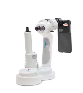
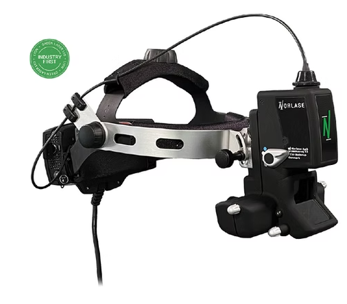
Muscat, Oman
+919359902383
+919359902383

ophthalmic devices

6814772a5d263414004dd4a4 Card 2

product image
Ophthalmic Devices

The Norlaseยฎ LIONโ„ข is a green laser indirect ophthalmoscope designed with you and your patients in mind. LIONโ€™s ultra-compact laser technology is built-in and powered by a battery in the footswitch โ€“ allowing doctors to enjoy an untethered, lightweight and portable laser treatment solution. A wireless user interface and voice control of parameters provide unmatched ease of use and practice efficiency. LION Redefining Laser Photocoagulator Treatment REINVENT YOUR PRACTICE Free from a cumbersome external fiber optic cable, LIONโ€™s integrated and untethered design allows you to treat patients anytime, anywhere. LION is the new gold standard LIO, combining functionality and comfort with reliability, unmatched ease of use, and portability. LION Designed with You and Your Patients in Mind BRING LION TO YOUR PRACTICE LASER SOURCE IN THE HEADSET High power, miniaturized green laser is smaller than a coin and fits seamlessly in the headset, eliminating the need for an external laser console. LION provides an all-in-one treatment solution that allows for unprecedented portability. SUPERIOR OPTICS Ultra-clear optics enhance visualization during diagnosis and treatment allowing physicians to treat faster and more efficiently. NO FIBER TETHER Laser-integrated headset with built-in fiber optics lets you move freely, minimizes disruptive service repairs, all while maximizing practice efficiency. BATTERY-POWERED High-capacity battery in the footswitch provides days of use between charges. No need to find a power outlet โ€“ just bring LION to your patient and start treating. WIRELESS TECHNOLOGY Parameter settings are fully operable through an intuitive user interface on a sleek wireless tablet. INDUSTRY-FIRST VOICE CONTROL Sophisticated speech recognition capability provides convenient voice control of laser parameters allowing you to stay focused on your patients. PORTABILITY AND PRACTICE EFFICIENCY Treat your patients anywhere, anytime. Unlike other LIOs, LIONโ€™s truly untethered design provides greater portability and flexibility so you can focus on whatโ€™s important โ€“ providing quality laser treatments that your patients are seeking.

6814772a5d263414004dd4a4 Card 2

product image
Ophthalmic Devices

Compact, versatile slit lamp with desktop operability.The SL-17 weighs less than 800g and provides easy and versatile operation with a choice of 10x and 16x magnifications, 3 slit widths* (0.1mm, 0.2mm, 0.8mm) plus spot illumination and a cobalt blue filter using a high illumination LED as the light source. The cobalt blue filter is useful for foreign body and contact lens applications. The SL-17 has a rheostat controlled light source from 0 to 20,000 Lux. The LED light source provides a much whiter light than ordinary halogen light sources found on competitive instruments. The SL-17 is compact and portable making it the ideal instrument for domiciliary environments, mobile evaluations, pediatrics, and patients not able to be imaged by a traditional slit lamp. The SL-17 is especially useful in emergency rooms where patient mobility is a challenge, or when switching between consultancy rooms is necessary. Offering cordless operation, the SL-17 is powered by (4) AAA rechargeable or dry cell batteries to provide long lasting, continuous operation of the extra-bright LED lamp. Features and Benefits 20,000 Lux White LED Light Source Illumination is thumb wheel controlled and illuminated for easy viewing in a darkened room. Powered by AAA Batteries The Kowa SL-17 uses the following commercially available batteries: AAA rechargeable batteries AAA dry cell batteries * Excludes lithium-ion and lithium batteries Anti-Tip Design New circular base increases slit lamp stability providing a safer place to store the instrument Available in 4 Colors

6814772a5d263414004dd4a4 Card 2

product image
Ophthalmic Devices

The next generation of pediatric ocular imaging. RetCam Envisionโ„ข sets the standard in pediatric ocular imaging, delivering brilliant images, seamless connectivity, and efficient workflow. RetCamยฎ Envision ophthalmic imaging system The next generation of pediatric ocular imaging. RetCamยฎ Envision sets the standard in pediatric ocular imaging, delivering brilliant images, seamless connectivity, and efficient workflow. The success of RetCam represented a huge leap in pediatric ocular imaging, setting a new standard in the field. With RetCam Envision Natus builds on that legacy of innovation and breaks new ground with creating exciting new developments in image quality, connectivity and workflow efficiency. Make a visible difference The new high-definition 80ยฐ magnification lens for RetCam Envision is an exciting new addition to the RetCam lens portfolio, embodying Natusโ€™ commitment to developing new and better state-of-the-art imaging options that make a visible difference to patient outcomes. Enhanced magnification Experience unparalleled clarity and detail with our 80ยฐ Field-of-View lens for magnified views of: Pediatric disease entities Macular disorders Vascular details/features Tumors and lesions Posterior pole External eye Brilliant imaging Enhanced illumination and ultra-wide field of view make RetCam Envision the leader in image quality for baby eye exams. These unique features result in images that provide a higher level of confidence for patient evaluations and consultations. Brilliant color images can be viewed in full-screen for maximum ophthalmic visualization of detail, early detection and timely intervention. Seamless connectivity RetCam Envision provides seamless PACS integration and enhanced network capabilities. Images are securely stored centrally within PACS, significantly improving efficiency by reducing patient intake time with manual data transfer and image archiving.* Optimal efficiency RetCam Envision is a perfect example of advanced technology meets practical application. This advance in pediatric ophthalmic equipment is easy to use and integrate into any NICU environment, and gives NICU staff control of the screening process and the ability to schedule eye exams based on their knowledge of the patientโ€™s status. Designed for accuracy and convenience Detachable 130ยฐ lens, and optional portrait lens provides greater visualization with brilliant color images with RetCam Envision Light Shaping Technology that can be viewed in full-screen mode on an expansive 21.5โ€ณ monitor. The lens can be removed easily for cleaning and disinfecting with minimal downtime. RetCam's Light Shaping Technology RetCam Envisionโ€™s innovative Light Shaping Technology provides uniform illumination for luminous detail in captured images. It provides focus across the entire 130ยบ field of view, improved peripheral illumination, and better illumination of darkly pigmented retinas. Fluorescein angiography delivers the crucial information you need Fluorescein angiography (FA) helps deliver essential information you need. RetCam Envisionโ€™s fluorescein angiography option is a diagnostic aid to examine the circulation of the retinal blood vessels and assist with the detection of anatomical abnormalities.* It provides users with the ability to visualize the absence or presence of blood flow within the eye. Benefits of FA: Helps to examine the perfusion of the retinal blood vessels Provides sharp visualization of leaking vessel defects and neovascularization in ROP Documents and supports the diagnosis of many pediatric ophthalmic diseases and conditions *Available for purchase separately Innovative tool for seamless integration Introducing DataLink for RetCam Envision, an innovative cloud-based platform that manages pediatric ocular exam images and data. DataLink works in tandem with RetCam Envision to offer remote viewing, advanced interpretation, communication and reporting capabilities. Optimize your clinical workflow with this secure and convenient centralized solution.

6814772a5d263414004dd4a4 Card 2

product image
Ophthalmic Devices

The All-New, Blazing Fast, Fully-Automated Specular Microscope > Widescreen Touch Panel PC mounted > Fully automated, integrated database > Location specific data samples > Robust Analysis Methods CellChekยฎ๏ธ๏ธ๏ธ 20 & 20 PLUS The All-New, Blazing Fast, Fully-Automated Specular Microscopes Process bilateral exams with one touch in about 40 seconds Konan specular microscopes were recognized as the gold standard specular microscopes in the minutes from an FDA panel meeting* The Gold Standard, Redefined. Faster, Easier Endothelial Imaging CellChekยฎ๏ธ๏ธ๏ธ 20 is Konan Medicalโ€™s new, non-contact specular microscope that can capture and analyze bilateral exams with one touch in under 40 seconds. The new easy-to-use โ€œSimple Modeโ€ enables one-touch, fully-automated endothelial image capture with analysis, reporting, and exporting of data. ๏ƒพ Clinical Applications Glaucoma, cataract, & refractive surgery. Corneal disease management. Contact/specialty contact lens fittings. Routine eye care N Clinical Benefits Visualize endothelial cells with 40x magnification compared to slit lamp bio-microscopy Identify pre-existing low density and dystrophies that may affect positive surgical outcomes Confirm recommended minimum cell density/morphology for scleral/specialty contact lenses ๏™ฏ Regulatory FDA 510(k) Cleared | CPT Code 92286 CE Marked Health Canada Licensed Blazing Fast The new, easy-to-use โ€œSimple Modeโ€ enables one-touch, fully-automated endothelial image capture with analysis, reporting, and exporting of data. The completely reimagined CellChekยฎ๏ธ๏ธ๏ธ 20 is noticeably improved and simplified; remove the device from the box, connect to power, turn it on, and start imaging. Konanโ€™s gold standard Center Methodโ„ข and Flex Center Methodโ„ข are now fully-automated. Automated Center Method displays cell center counted densities and morphometric indices of both eyes immediately after images are captured โ€“ all in under 40 seconds. Key New Features With CellChekยฎ๏ธ๏ธ๏ธ 20 you can capture, analyze and print/export a bilateral exam with just one touch. The new, easy-to-use โ€œSimple Modeโ€ makes running a bilateral exam faster and easier than ever before. CellChekยฎ๏ธ๏ธ๏ธ 20 reduces testing time significantly, allowing you to test patients quickly and efficiently. ๏‚ฆ One-touch imaging Fast, one-touch imaging of both eyes including positioning, alignment, imaging capture, analysis and print-out/exporting data. ๏…ค Optimized touch functions Easy touch-friendly interface makes capturing, analysis, and reporting fast and efficient. Also features pinch-to-zoom image adjustment. ๏‚ฆ 25% higher resolution Improved resolution allows better visualization of single cells and cell walls. Superb cell border tracing and guttae exclusion. ๏€ก Auto-capture/retry This improved technology allows for successful imaging of difficult or challenging cases. ๏Œž 30% larger image area The field of view has been increased to 0.25mm x 0.55mm, enabling better corneal visualization and more cells to be analyzed. CellChekยฎ๏ธ๏ธ๏ธ 20 PLUS CellChekยฎ๏ธ๏ธ๏ธ 20 PLUS adds near-limbal imaging to the standard model. It extends the limits of corneal endothelial image capture adding locations to 4.5mm from center. Six additional fixation points, 13 total. Flexible Touch Screen The flexible 10.6โ€ wide touch screen can be turned and tilted 180ยฐ, providing better ergonomics, space savings, and flexibility with technician use from any of the four sides: front, back, left, and right. Fully-Automated Analysis CellChekยฎ๏ธ๏ธ๏ธ 20 offers fully-automated: Center Method; Flex-Center Method; and Auto-Trace analysis. These analysis methods provide one-touch, high precision results FAST. These fully-automated analysis methods are offered alongside the traditional manual Center and Flex-Center analysis methods, offering a total of 6 analysis options. Konanโ€™s Center Method is mentioned in FDA panel minutes as being the โ€œgold standardโ€, and is used by virtually every professional reading center. New Auto Center Method With the new Auto Center Method, the center of each individual, visible cell is automatically marked. Guttae and other dark regions are automatically excluded. New Auto Flex-Center Method With the new Auto Center Method, the center of each individual, visible cell is automatically marked. Guttae and other dark regions are automatically excluded. Clinical Benefits Cataract Surgery and Premium IOLs Low endothelial cell counts and pre-existing dystrophies can markedly reduce the potential for positive surgical outcomes from an otherwise uneventful cataract surgery. Surgeons are finding these data points critical when recommending premium IOLs: verify and document pre-operatively that the cornea is not suspect to more likely post-op complications difficult to explain with the investment in premium IOLs.

6814772a5d263414004dd4a4 Card 2

product image
Ophthalmic Devices

CellChekยฎ D | D+ Eye Bank Specular Microscopes Designed to improve patient outcomes, CellChekยฎ D+ redefined specular microscopy in Eyebanking. It was quickly and widely adopted by many of the worldโ€™s finest Eye Banks. CellChekยฎ D+ Introducing the first multi-imaging system for donor corneal analysis. CellChekยฎ D+ provides an amazing view into the cornea that simply has never been seen before defining the structure of the endothelium, stroma and epithelium. Comprehensive imaging to assist both eye banks and corneal surgeons to make better decisions on implantable material. Now featured in Cornea June 2018 Supplement: Current and New Technologies in Corneal Donor Tissue Evaluation Comparative Image Atlas. Especially check out S7 โ€œWide-Field Ex Vivo Dual Imaging Microscopy.โ€ Clinical Benefits Provides remarkable views of endothelium, intra-stomal structure and the epithelium For the first time, visualize blood cells, fungus, rough keratome cuts, dead cells, and epithelial anomalies New โ€œDonor Enhancedโ€ Imaging System CellChekยฎ D+ features a patent applied for โ€œDonor Enhanceโ€ imaging system that provides remarkable views of endothelium, intra-stomal structure, and the epithelium. The range of new information from which to make better clinical decisions is spectacular (pun intended): blood cells, fungus, rough keratome cuts, dead cells, epithelial anomalies. Never have we had this view of pre-implanted material. Corneal surgeons are finding this information to be illuminating in helping to understand graft success and failure. Broad overview but with large multi-sample analytic areas CellChekยฎ D and D+ feature a new finder feature with digital measuring / documenting tools. Patented Cellular Analysis Methods All Konan specular microscopes feature the Center Method of analysis. Center Method is mentioned in FDA panel minutes as being the โ€œgold standardโ€ and is used by virtually every professional reading center for independent assessment of corneal endothelial analytics.ยน Unlike full-auto assessments, the Center Method provides high precision and repeatability for specular images in which relatively small continuous areas of visible cells are visible. The Flex-Center method is the third tool for advanced stage diseased corneas in which only a very few number of cells are visible. With this semi-automated, perimeter-count method, again, high precision and repeatability is achieved. Only Konan provides the rich set of analytic tools for reliable assessment of the entire spectrum of corneal conditions. It is not surprising that Konan is the global gold standard in corneal endothelial assessment. Full Graft Imaging CellChekยฎ D and D+ provide a total picture of the cornea. Use digital measurement tools to identify, measure and document cornea dimensions, defects, and scars.

6814772a5d263414004dd4a4 Card 2

product image
Ophthalmic Devices

Innovative Cyclophotocoagulation with MicroPulseยฎ Technology Powered by the New CYCLO G6โ„ข Glaucoma Laser System. Reduces IOP by 30-50% in a safe tested, repeatable procedure and lower the burden of numerous glaucoma medications for your patients. For MicroPulse transscleral laser therapy and Continuous-Wave Cyclophotocoagulation Can be used in the office and operating room Used in 38 of the 39 best U.S. hospitals Available in over 80 countries

6814772a5d263414004dd4a4 Card 2

product image
Ophthalmic Devices

EndoProbe instrumentation targets the retina to deliver precise energy exactly where you need it. With a wide array of models, there is an EndoProbe for every vitreoretinal laser case. Extended Reach. Enhanced Performance. Excellent uptake at varied distances from the target tissue Easy, smooth extension and retraction of probe Compatible with valved cannulas Adjustable & Intuitive (Finger or Thumb) Patented design allows continuous adjustment of fiber optic over a wide range of angles Provides full coverage of peripheral retina without removing probe from eye Extends in logical motion, forming a greater angular deflection as slider is advanced Available in 20G, 23G, and 25G in finger or thumb adjustable models Stepped Angled Smooth and gently tapered needle that permits insertion of angled tip through standard and valved cannulas Patented design provides full coverage of peripheral retina without removing probe from eye Offers rigid, fixed angle for a sturdy alternative to flex-tip probes Laser probes available in 20G, 23G, and 25G models Illuminating Laser Probes Dual function - white-light illumination with laser delivery in one convenient design Offers bimanual operation - one hand manages illumination and laser delivery, freeing the other hand to operate additional instruments Optimal brightness - Combines multiple illumination fibers and one laser fiber Available in 19.5G, 20G, 23G, and 25G models Standard Straight Provide direct access to treatment site Facilitate easy insertion and extraction at the sclerotomy site Allow easier insertion and visibility due to tapered tip Available in 20G, 23G, and 25G models Standard Angled Used for treatment of the peripheral retina Provides greater flexibility when using a wide field viewing system Includes a tapered tip for easier insertion and visibility of the treatment area Available in 20G model Active Aspirating Combines the utility of active aspiration and endophotocoagulation in a single device Eliminates the need for extrusion needles and frees hand for illumination Includes Luer fitting compatible with standard aspirating equipment Available in 20G model Passive Fluted Aspirating For subretinal fluid aspiration associated with tears and detachments Combines the utility of passive aspiration and endophotocoagulation in a single device Designed for surgeons who prefer to control the rate of fluid extrusion with their finger Available in 20G model

6814772a5d263414004dd4a4 Card 2

product image
Ophthalmic Devices

MicroPulseยฎ Laser Therapy for repeatable, tissue-sparing* glaucoma and retinal therapies. PRODUCT HIGHLIGHTS High power for greater range of therapy alternatives Dual port for efficient setup of alternate delivery devices Repeatable laser sessions for retinal diseases with optional MicroPulse module The Advantages of Innovation High power for greater range of therapy alternatives High speed pulse durations for faster procedural time DualSense provides quick and simple selection of multiple delivery devices for RFID and SMA Intelligent and intuitive design Dual Port for efficient setup of alternate delivery devices Intuitive graphic touch screen interface for ease of use Programmable memory presets for multiple users Optional Module for MicroPulse Laser Therapy, Fovea-Friendly* Repeatable laser sessions for retinal diseases Repeatable glaucoma therapy with MicroPulse laser trabeculoplasty (MLT) IQ 532ยฎ Laser System Wavelength 532 nm Green Weight 19.2 lb (9.0 kg) Dimensions 8.5โ€œ H x 12โ€œ W x 14โ€œ D (21 cm x 30 cm x 35 cm) Connector Type RFID | Resistor Electrical 100-240 VAC, 50/60 Hz Cooling Whisper fan Exposure Duration CW-Pulse: 10-3000 ms Exposure Interval CW-Pulse: 10-3000 ms MicroPulse Duration MicroPulse: 0.05-1.00 ms MicroPulse Interval MicroPulse: 1.00-10.00 ms Aiming Laser Diode laser, 635 nm nominal Delivery Device Power Output Portable SLA: 0-2000 mW LIO: 0-2000 mW EndoProbe: 0-2000 mW Optional Accessories Full-Featured Remote Control Compact design for easy placement or use in sterile field View displays and adjust parameters from two vantage points for increased convenience and efficiency Wireless Footswitch No cord, no clutter, no limitations Allows precise physician control over power settings Available with power-adjust to control laser actuation and power settings

6814772a5d263414004dd4a4 Card 2

product image
Ophthalmic Devices

TxCellโ„ข Scanning Laser Delivery System Versatile Platform for Advanced Retinal Therapy The TxCell Scanning Slit Lamp Adapter adds the use of multi-spot pattern scanning when coupled to the IQ 532TM or IQ 577TM laser. In one platform, growing practices can offer: ยฐ Multi-spot pattern scanning for efficient pan retinal photocoagulation ยฐ Standard photocoagulation with optimized wavelengths: IQ 532 and true-yellow IQ 577 ยฐ Confluent laser patterns for tissue-sparing MicroPulseTM protocols Workflow Efficiency ยฐ Predictability in laser spot placement for both standard photocoagulation and MicroPulse protocols ยฐ High speed pulse durations for efficient laser delivery ยฐ Modular design for intra-office portability IQ 532TM and IQ 577TM Laser Systems The Advantages of Innovation ยฐ High power for greater range of therapy alternatives ยฐ High speed pulse durations for faster procedural time ยฐ DualSenseTM provides quick and simple selection of multiple delivery devices for RFID and SMA Intelligent and Intuitive Design ยฐ Dual port for efficient setup of alternate delivery devices ยฐ Intuitive graphic touch screen interface for ease of use ยฐ Programmable memory presets for multiple users TxCellโ„ข Scanning Laser Delivery System Specifications are subject to change without notice. IRIDEX and the IRIDEX logo are registered trademarks and IQ 532, IQ 577, DualSense, CW-Pulse, MicroPulse and Fovea-Friendly are trademarks of IRIDEX Corporation. All other trademarks are the property of their respective owners. Products are covered by one or more of the following U.S. patents: 5,372,595; 5,511,085; 5,521,932; 5,663,979; 5,982,789; 5,979,554; 6,141,143; 6,144,484; 6,222,869; 6,327,291; 6,377,599; 6,540,391; 6,733,490; 6,800,076; 7,537,593; 7,766,904; 7,771,417; and 7,909,816. LASER RADIATION AVOID EYE OR SKIN EXPOSURE TO DIRECT OR SCATTERED RADIATION CLASS 4 LASER PRODUCT (IEC 60825-1:2007) LASER RADIATION DO NOT STARE INTO BEAM CLASS 2 LASER PRODUCT ฮป = 532 nm PO = 5 W ฮป = 577 nm PO = 5 W ฮป = 635 nm PO = 2 mW EC REP Specifications Laser: IQ 532โ„ข/IQ 577โ„ข Wavelength: 577 nm or 532 nm Laser Energy Source: Frequency doubled solid-state and direct diode Maximum Power: 2 W Exposure Duration: CW-Pulseโ„ข: 10โ€“3000 ms Exposure Interval: CW-Pulse: 10โ€“3000 ms MicroPulseโ„ข Duration: MicroPulse: 0.05โ€“1.00 ms MicroPulse Interval: MicroPulse: 1.00โ€“10.00 ms Aiming Beam: Diode laser, 635 nm nominal Patterns: Grid (2x2 - 7x7), Circle, Triple Arc User Interface: Touchscreen & knobs Slit Lamp: CSO SL 980, Zeiss 30SL, Zeiss SL 130, and equivalents Spot Sizes: Single spot: 50ยตm, 100ยตm, 200ยตm, 300ยตm, 500ยตm Multi-spot: 100ยตm, 200ยตm, 300ยตm, 500ยตm Electrical: 100 โ€“ 240 VAC, 50/60 Hz The TxCell Scanning Laser Delivery System components: ยฐ TxCell Scanning Slit Lamp Adapter (SSLA) ยฐ TxCell Control Box ยฐ IQ 532 or IQ 577 laser

6814772a5d263414004dd4a4 Card 2

product image
Ophthalmic Devices

MicroPulseยฎ Laser Therapy for Fovea-Friendlyโ„ข* laser grids and tissue-sparing* combo therapies. PRODUCT HIGHLIGHTS High transmission through dense ocular media Dual Port for efficient setup of alternate delivery devices Repeatable macular and foveal laser sessions Maximize workflow efficiency True Yellow, 577nm wavelength for precise performance High transmission through dense ocular media Consistent laser lesions for fast procedure time Low power required for increased patient comfort Intelligent and intuitive design Dual Port for efficient setup of alternate delivery devices Intuitive graphic touch screen interface for ease of use Programmable memory presets for multiple users Optional Module for MicroPulse Laser Therapy Repeatable macular and foveal laser sessions Success with refractory and sub-clincal edema IQ 577ยฎ Laser System Wavelength 577 nm Yellow Weight 19.2 lb (9.0 kg) Dimensions 8.5โ€œ H x 12โ€œ W x 14โ€œ D (21 cm x 30 cm x 35 cm) Connector Type RFID | Resistor Electrical 100-240 VAC, 50/60 Hz Cooling Whisper fan Exposure Duration CW-Pulse: 10-3000 ms Exposure Interval CW-Pulse: 10-3000 ms MicroPulse Duration MicroPulse: 0.05-1.00 ms MicroPulse Interval MicroPulse: 1.00-10.00 ms Aiming Laser Diode laser, 635 nm nominal Delivery Device Power Output Portable SLA: 0-2000 mW LIO: 0-2000 mW EndoProbe: 0-2000 mW Optional Accessories Full-Featured Remote Control Compact design for easy placement or use in sterile field View displays and adjust parameters from two vantage points for increased convenience and efficiency Wireless Footswitch No cord, no clutter, no limitations Allows precise physician control over power settings Available with power-adjust to control laser actuation and power settings

6814772a5d263414004dd4a4 Card 2

product image
Ophthalmic Devices

The OcuLightยฎ Tx Green 532nm Laser is designed with higher power for conventional ophthalmic and otolaryngological procedures. Combining high power with high speed, the device offers superb efficiency with a full 2500 mW of deliverable laser energy. And it provides pulse durations and pulse intervals from 10 to 3000 milliseconds, using TrueCWโ„ข laser pulses for maximum control and precision. The expandable system offers a broad selection of EndoProbeยฎ and OtoProbeโ„ข handpieces for a variety of surgical procedures, as well as optional remote control and wireless footswitch. It is also ergonomic and truly portable, transporting easily between treatment rooms and offices. Features Includes TrueCWโ„ข laser pulses for maximum control and precision Designed for durability, with a thermal management system to increase laser conversion efficiency and reliability Enhances user interface with conveniently located, easily understood controls Allows easy transport between rooms and offices PowerSuperb efficiency with a full 2500 mW of deliverable laser energy. Delivery SystemIncludes a full line of delivery devices. Weight (lbs)13.2 lbs (<6.0 kg)> Power Input100โ€“240 VAC, 50/60 Hz Dimensions (W ร— H ร— D)12 in (30 cm) x 6 in (15 cm) x 12 in (30 cm) Aim BeamDiode laser, 635 nm nominal Wavelength532 nm CoolingWhisper fan with peltier cooling

6814772a5d263414004dd4a4 Card 2

product image
Ophthalmic Devices

The Octopus 600 offers standard central field white-on-white perimetry and fast screening in one compact and dark room- independent device. It includes easy-to-read and sensitive progression analysis and networking capability, thanks to its integrated EyeSuite software. Central field static perimetry Covering all essential needs The Octopus 600 offers a comprehensive library of standard central visual field tests. It includes the G, M, 24-2, 24-2C, 30-2, 32 and 10-2 patterns and tests a full threshold in just 2-4 minutes with the TOP strategy. Pulsar and screening Screening in typically less than one minute The Octopus 600 allows for fast screening and a quick distinction between normal and abnormal visual fields. Screening can be performed with both standard white-on-white or the patient-friendly Pulsar perimetry designed for early glaucoma detection. Octopus & HFA view Flexibility for your practice Easily switch to HFA mode and view your single fields, progression, and even printouts in the familiar Humphrey format. Whether you need to exchange data with colleagues or take advantage of the best of both worlds, the Octopus perimeter offers full flexibility in data analysis. Perimetry simplified Usability & ergonomics The compact Octopus 600 fits almost anywhere and can be operated reliably outside of a darkroom. Its design supports ergonomics positioning for the operator and the patient. EyeSuite Integration into practice workflows Our EyeSuite software has been designed to support patient flow in busy practices. It controls many Haag-Streit devices and allows for them to be easily networked, whether that's with other Haag-Streit devices, an office computer, or an EMR systemโ€” all without the need for any third-party software. EyeSuite glaucoma analysis View glaucoma from different angles The Octopus 600 allows you to view glaucoma visual field series form different angles. Take a look at the highly sensitive Cluster Analysis & the Polar Analysis for structural comparision for single field and progression. Explore our educational resources Find learning materials by product or category Access a variety of materials designed to help you get the most from our products. From demonstration videos and webinars to publications, studies, and best-practice documents โ€“ explore resources that provide practical guidance, application insights, and product knowledge, available whenever and wherever you choose to learn. Haag-Streit UK Service Division Let us service your perimeter An experienced and fully-trained Field Service Engineer from the HS-UK Service Division will visit your premises and carry out the following service procedures on your Octopus perimeter: 1. Check EyeSuite version and that the system operates without errors 2. Clean all optics and mirrors 3. Carry out a perimetry test 4. Ensure chinrest operates freely and holder moves in and out of position 5. Ensure patient answering switch is operating correctly. At HS-UK, we only use genuine Haag-Streit approved parts, which ensures your Octopus device complies with Haag-Streit specifications.

Phone Number

+919405476170

Mon-Thu: 10 AM - 2 PM • Fri: 3 PM - 7AM

Other Website Visit our other website
Address Muscat, Oman

Muscat, Oman, Oman, 112